Grupo Inmunovirologia, Universidad de Antioquia, Medellín, Antioquia, Colombia.
PLoS One. 2012;7(1):e30307. doi: 10.1371/journal.pone.0030307. Epub 2012 Jan 19.
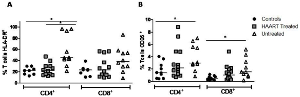
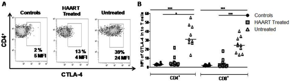
Peripheral blood T-cells from untreated HIV-1-infected patients exhibit reduced immune responses, usually associated with a hyperactivated/exhausted phenotype compared to HAART treated patients. However, it is not clear whether HAART ameliorates this altered phenotype of T-cells in the gastrointestinal-associated lymphoid tissue (GALT), the main site for viral replication. Here, we compared T-cells from peripheral blood and GALT of two groups of chronically HIV-1-infected patients: untreated patients with active viral replication, and patients on suppressive HAART. We characterized the T-cell phenotype by measuring PD-1, CTLA-4, HLA-DR, CD25, Foxp3 and granzyme A expression by flow cytometry; mRNA expression of T-bet, GATA-3, ROR-γt and Foxp3, and was also evaluated in peripheral blood mononuclear cells and rectal lymphoid cells. In HIV-1+ patients, the frequency of PD-1(+) and CTLA-4(+) T-cells (both CD4+ and CD8+ T cells) was higher in the GALT than in the blood. The expression of PD-1 by T-cells from GALT was higher in HIV-1-infected subjects with active viral replication compared to controls. Moreover, the expression per cell of PD-1 and CTLA-4 in CD4(+) T-cells from blood and GALT was positively correlated with viral load. HAART treatment decreased the expression of CTLA-4 in CD8(+) T cells from blood and GALT to levels similar as those observed in controls. Frequency of Granzyme A(+) CD8(+) T-cells in both tissues was low in the untreated group, compared to controls and HAART-treated patients. Finally, a switch towards Treg polarization was found in untreated patients, in both tissues. Together, these findings suggest that chronic HIV-1 infection results in an activated/exhausted T-cell phenotype, despite T-cell polarization towards a regulatory profile; these alterations are more pronounced in the GALT compared to peripheral blood, and are only partiality modulated by HAART.
未经治疗的 HIV-1 感染患者的外周血 T 细胞表现出免疫反应降低,通常与接受高效抗逆转录病毒治疗(HAART)的患者相比具有高激活/耗竭表型。然而,尚不清楚 HAART 是否能改善胃肠道相关淋巴组织(GALT)中 T 细胞的这种改变表型,GALT 是病毒复制的主要部位。在这里,我们比较了两组慢性 HIV-1 感染患者的外周血和 GALT 中的 T 细胞:未经治疗且病毒复制活跃的患者和接受抑制性 HAART 的患者。我们通过流式细胞术测量 PD-1、CTLA-4、HLA-DR、CD25、Foxp3 和颗粒酶 A 的表达来表征 T 细胞表型;还评估了外周血单核细胞和直肠淋巴细胞中 T-bet、GATA-3、ROR-γt 和 Foxp3 的 mRNA 表达。在 HIV-1+ 患者中,GALT 中 PD-1(+)和 CTLA-4(+)T 细胞(CD4+和 CD8+T 细胞)的频率高于血液。与对照相比,具有活跃病毒复制的 HIV-1 感染患者的 GALT 中 T 细胞表达 PD-1 更高。此外,血液和 GALT 中 CD4(+)T 细胞中 PD-1 和 CTLA-4 的每个细胞表达与病毒载量呈正相关。HAART 治疗将血液和 GALT 中 CD8(+)T 细胞中 CTLA-4 的表达降低至与对照相似的水平。与对照和 HAART 治疗的患者相比,未经治疗组的两种组织中 CD8(+)T 细胞中颗粒酶 A(+)的频率较低。最后,在未经治疗的患者中,两种组织中均发现 Treg 极化向调节表型的转变。总之,这些发现表明,慢性 HIV-1 感染导致激活/耗竭的 T 细胞表型,尽管 T 细胞向调节表型极化;与外周血相比,这些改变在 GALT 中更为明显,并且仅部分被 HAART 调节。