Department of Nephrology, The First Affiliated Hospital, Sun Yat-sen University, Key Laboratory of Nephrology, Ministry of Health, Guangzhou, Guangdong, China.
PLoS One. 2012;7(1):e30312. doi: 10.1371/journal.pone.0030312. Epub 2012 Jan 20.
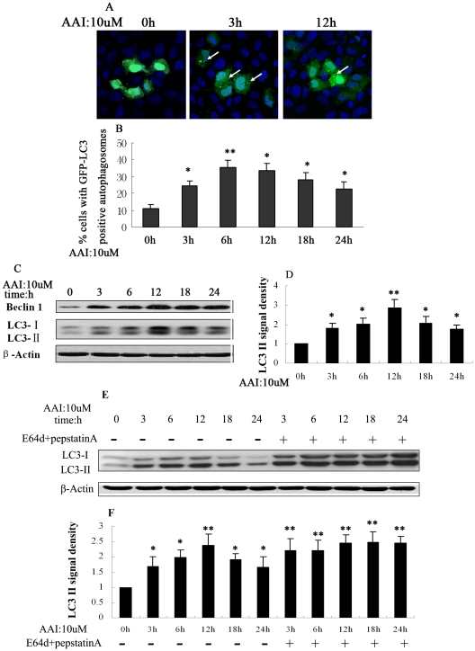
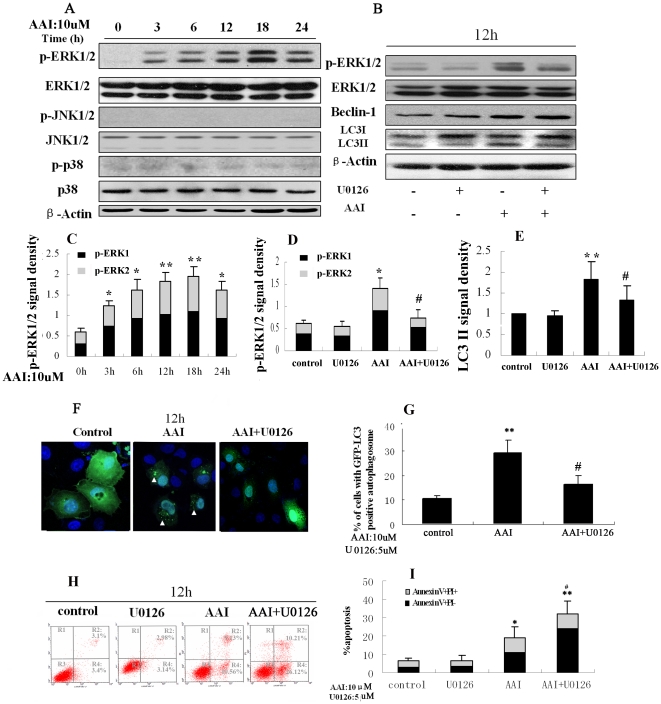
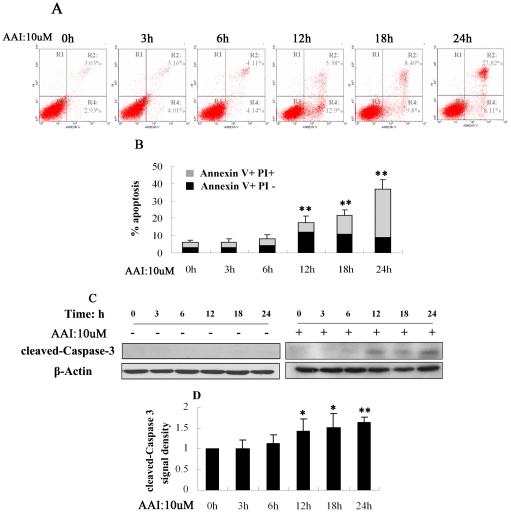
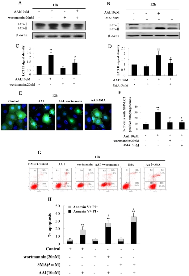
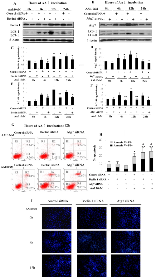
Autophagy is a lysosomal degradation pathway that is essential for cell survival and tissue homeostasis. However, limited information is available about autophagy in aristolochic acid (AA) nephropathy. In this study, we investigated the role of autophagy and related signaling pathway during progression of AAI-induced injury to renal tubular epithelial cells (NRK52E cells). The results showed that autophagy in NRK52E cells was detected as early as 3-6 hrs after low dose of AAI (10 µM) exposure as indicated by an up-regulated expression of LC3-II and Beclin 1 proteins. The appearance of AAI-induced punctated staining of autophagosome-associated LC3-II upon GFP-LC3 transfection in NRK52E cells provided further evidence for autophagy. However, cell apoptosis was not detected until 12 hrs after AAI treatment. Blockade of autophagy with Wortmannin or 3-Methyladenine (two inhibitors of phosphoinositede 3-kinases) or small-interfering RNA knockdown of Beclin 1 or Atg7 sensitized the tubular cells to apoptosis. Treatment of NRK52E cells with AAI caused a time-dependent increase in extracellular signal-regulated kinase 1 and 2 (ERK1/2) activity, but not c-Jun N-terminal kinase (JNK) and p38. Pharmacological inhibition of ERK1/2 phosphorylation with U0126 resulted in a decreased AAI-induced autophagy that was accompanied by an increased apoptosis. Taken together, our study demonstrated for the first time that autophagy occurred earlier than apoptosis during AAI-induced tubular epithelial cell injury. Autophagy induced by AAI via ERK1/2 pathway might attenuate apoptosis, which may provide a protective mechanism for cell survival under AAI-induced pathological condition.
自噬是一种溶酶体降解途径,对细胞存活和组织稳态至关重要。然而,关于马兜铃酸(AA)肾病中的自噬知之甚少。在这项研究中,我们研究了自噬及其相关信号通路在 AAI 诱导的肾小管上皮细胞(NRK52E 细胞)损伤进展中的作用。结果表明,低剂量 AAI(10 μM)暴露后 3-6 小时,NRK52E 细胞中的自噬就被检测到,LC3-II 和 Beclin 1 蛋白的表达上调。在 GFP-LC3 转染的 NRK52E 细胞中,AAI 诱导的自噬体相关 LC3-II 点状染色的出现进一步证明了自噬的存在。然而,直到 AAI 处理 12 小时后才检测到细胞凋亡。用 Wortmannin 或 3-甲基腺嘌呤(两种磷酸肌醇 3-激酶抑制剂)或 Beclin 1 或 Atg7 的小干扰 RNA 敲低阻断自噬,使肾小管细胞对凋亡敏感。AAI 处理 NRK52E 细胞会导致细胞外信号调节激酶 1 和 2(ERK1/2)活性的时间依赖性增加,但不会导致 c-Jun N 末端激酶(JNK)和 p38 增加。用 U0126 抑制 ERK1/2 磷酸化可导致 AAI 诱导的自噬减少,同时凋亡增加。总之,我们的研究首次表明,在 AAI 诱导的肾小管上皮细胞损伤中,自噬发生早于凋亡。AAI 通过 ERK1/2 通路诱导的自噬可能会减轻凋亡,这可能为 AAI 诱导的病理条件下细胞存活提供一种保护机制。