Department of Physics and Astronomy, College of Science, King Saud University, Saudi Arabia.
J Nanobiotechnology. 2012 Jan 25;10:5. doi: 10.1186/1477-3155-10-5.
Nanoparticles (NPs) can potentially cause adverse effects on organ, tissue, cellular, subcellular and protein levels due to their unusual physicochemical properties. Advances in nanotechnology have identified promising candidates for many biological and biomedical applications. Since the properties of NPs differ from that of their bulk materials, they are being increasingly exploited for medical uses and other industrial applications. The aim of the present study was to investigate the particle-size effect of gold nanoparticles (GNPs) on the hepatic tissue in an attempt to cover and understand the toxicity and the potential threat of their therapeutic and diagnostic use.
To investigate particle-size effect of GNPs on the hepatic tissue, a total of 70 healthy male Wistar-Kyoto rats were exposed to GNPs received 50 or 100 ul of GNPs infusion of size (10, 20 and 50 nm for 3 or 7 days).
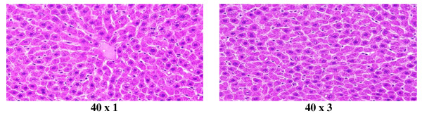
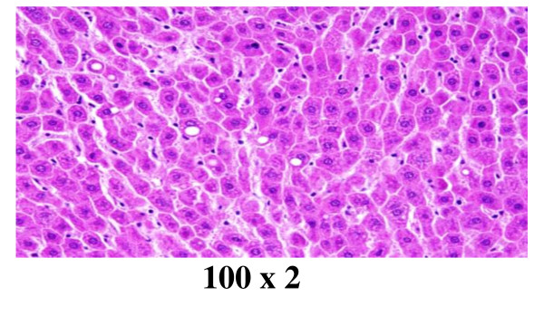
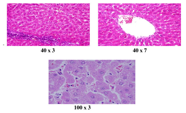
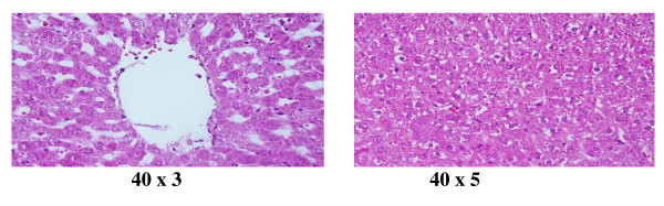
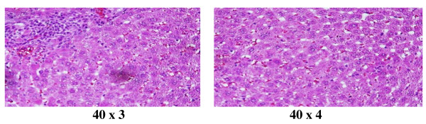
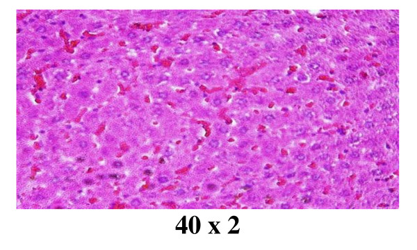
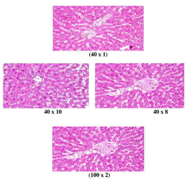
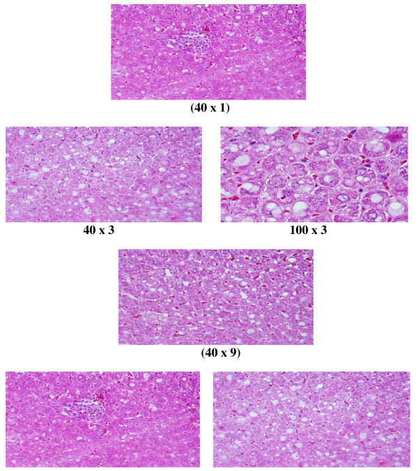
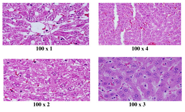
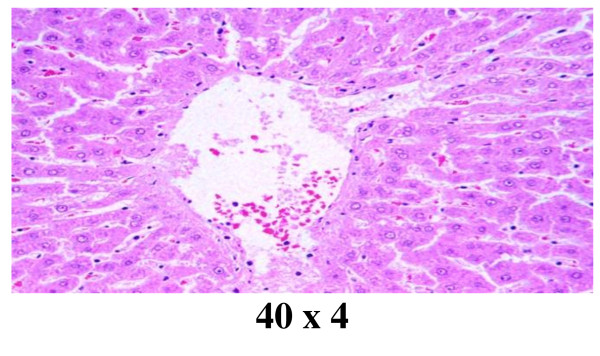
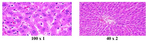
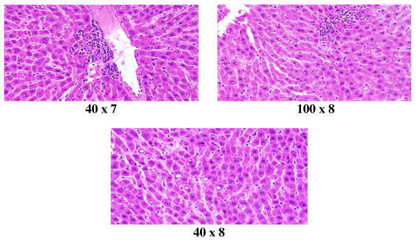
In comparison with respective control rats, exposure to GNPs doses has produced alterations in the hepatocytes, portal triads and the sinusoids. The alterations in the hepatocytes were mainly summarized as hydropic degeneration, cloudy swelling, fatty degeneration, portal and lobular infiltrate by chronic inflammatory cells and congestive dilated central veins.
The induced histological alterations might be an indication of injured hepatocytes due to GNPs toxicity that became unable to deal with the accumulated residues resulting from metabolic and structural disturbances caused by these NPs. These alterations were size-dependent with smaller ones induced the most effects and related with time exposure of GNPs. The appearance of hepatocytes cytoplasmic degeneration and nuclear destruction may suggest that GNPs interact with proteins and enzymes of the hepatic tissue interfering with the antioxidant defense mechanism and leading to reactive oxygen species (ROS) generation which in turn may induce stress in the hepatocytes to undergo atrophy and necrosis. More histomorphologcal, histochemical and ultrastrucural investigations are needed in relation of the application of GNPs with their potential threat as a therapeutic and diagnostic tool.
由于其异常的物理化学性质,纳米粒子(NPs)可能会对器官、组织、细胞、亚细胞和蛋白质水平造成不良影响。纳米技术的进步已经确定了许多生物和生物医学应用的有前途的候选者。由于 NPs 的性质与它们的块状材料不同,因此它们越来越多地被用于医疗用途和其他工业应用。本研究的目的是研究金纳米粒子(GNPs)的粒径效应对肝组织的影响,试图涵盖并了解其治疗和诊断用途的毒性和潜在威胁。
为了研究 GNPs 的粒径效应对肝组织的影响,总共将 70 只健康雄性 Wistar-Kyoto 大鼠暴露于 GNPs 中,分别接受 50 或 100ul 的 GNPs 输注,大小为(10、20 和 50nm)持续 3 或 7 天。
与各自的对照大鼠相比,暴露于 GNPs 剂量会导致肝细胞、门三联体和窦状隙发生改变。肝细胞的改变主要总结为水样变性、混浊肿胀、脂肪变性、门管区和小叶浸润慢性炎症细胞和充血扩张的中央静脉。
这些诱导的组织学改变可能是由于 GNPs 毒性导致肝细胞受损的迹象,这些细胞无法处理由于这些 NPs 引起的代谢和结构紊乱而产生的积累残留物。这些改变与粒径有关,较小的粒径会产生最大的影响,并与 GNPs 的暴露时间有关。肝细胞细胞质变性和核破坏的出现可能表明 GNPs 与肝组织的蛋白质和酶相互作用,干扰抗氧化防御机制,并导致活性氧(ROS)的产生,从而可能导致肝细胞应激,发生萎缩和坏死。需要进行更多的组织形态学、组织化学和超微结构研究,以了解 GNPs 的应用及其作为治疗和诊断工具的潜在威胁。