Chan Rs, Kumar G, Abdullah Bjj, Ng Kh, Vijayananthan A, Mohd Nor H, Liew Yw
Department of Biomedical Imaging, Faculty of Medicine, University of Malaya, Kuala Lumpur, Malaysia.
Biomed Imaging Interv J. 2011 Apr;7(2):e12. doi: 10.2349/biij.7.2.e12. Epub 2011 Apr 1.
To optimize the delay time before the initiation of arterial phase scan in the detection of focal liver lesions in contrast enhanced 5 phase liver CT using the bolus tracking technique.
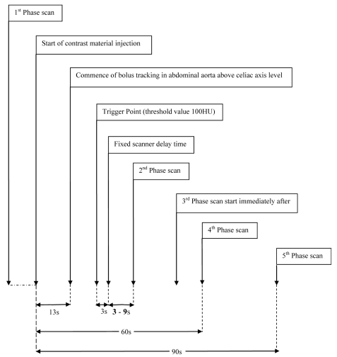
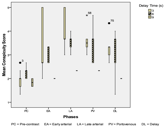
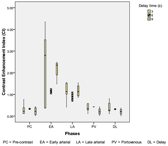
Delay - the interval between threshold enhancement of 100 hounsfield unit (HU) in the abdominal aorta and commencement of the first arterial phase scan. Using a 16 slice CT scanner, a plain CT of the liver was done followed by an intravenous bolus of 120 ml nonionic iodinated contrast media (370 mg I/ml) at the rate of 4 mL/s. The second phase scan started immediately after the first phase scan. The portal venous and delay phases were obtained at a fixed delay of 60 s and 90 s from the beginning of contrast injection. Contrast enhancement index (CEI) and subjective visual conspicuity scores for each lesion were compared among the three groups.
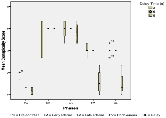
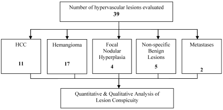
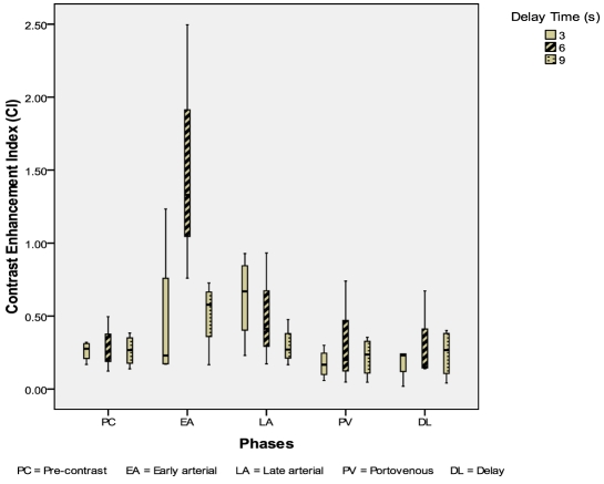
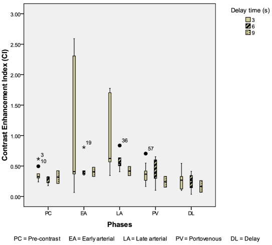
84 lesions (11 hepatocellular carcinomas, 17 hemangiomas, 39 other hypervascular lesions and 45 cysts) were evaluated. CEI for hepatocellular carcinomas appears to be higher during the first arterial phase in the 6 seconds delay group. No significant difference in CEI and mean conspicuity scores among the three groups for hemangioma, other hypervascular lesions and cysts.
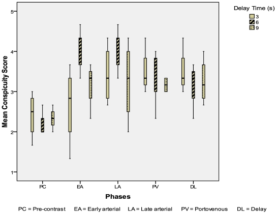
The conspicuity of hepatocellular carcinomas appeared better during the early arterial phase using a bolus tracking technique with a scan delay of 6 seconds from the 100 HU threshold in the abdominal aorta.
利用团注追踪技术优化肝脏CT增强扫描5期扫描中局灶性肝病变检测时动脉期扫描开始前的延迟时间。
延迟时间——腹主动脉强化达到100亨氏单位(HU)至首次动脉期扫描开始之间的间隔时间。使用16层CT扫描仪,先行肝脏平扫CT,然后以4ml/s的速率静脉团注120ml非离子型碘对比剂(370mg I/ml)。第一期扫描后立即开始第二期扫描。门静脉期和延迟期扫描在对比剂注射开始后分别固定延迟60秒和90秒进行。比较三组中每个病变的对比增强指数(CEI)和主观视觉清晰度评分。
共评估了84个病变(11个肝细胞癌、17个血管瘤、39个其他富血供病变和45个囊肿)。在延迟6秒的组中,肝细胞癌在第一动脉期的CEI似乎更高。血管瘤、其他富血供病变和囊肿在三组之间的CEI和平均清晰度评分无显著差异。
采用团注追踪技术,在腹主动脉100HU阈值时扫描延迟6秒,肝细胞癌在动脉早期的显示效果更佳。