Department of Anatomy and Structural Biology, Albert Einstein College of Medicine of Yeshiva University, 1300 Morris Park Avenue, Bronx, NY 10461, USA.
Traffic. 2012 May;13(5):643-9. doi: 10.1111/j.1600-0854.2012.01336.x. Epub 2012 Feb 20.
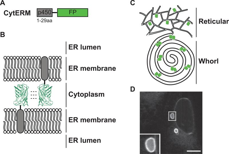
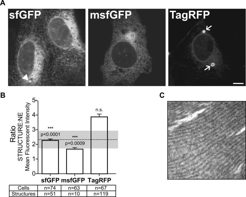
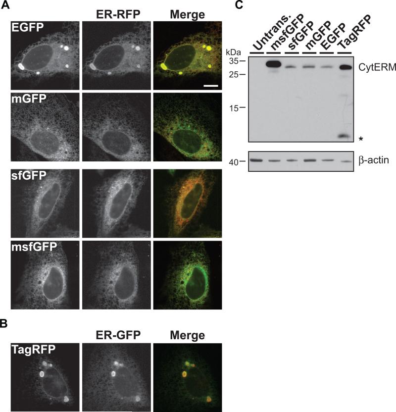
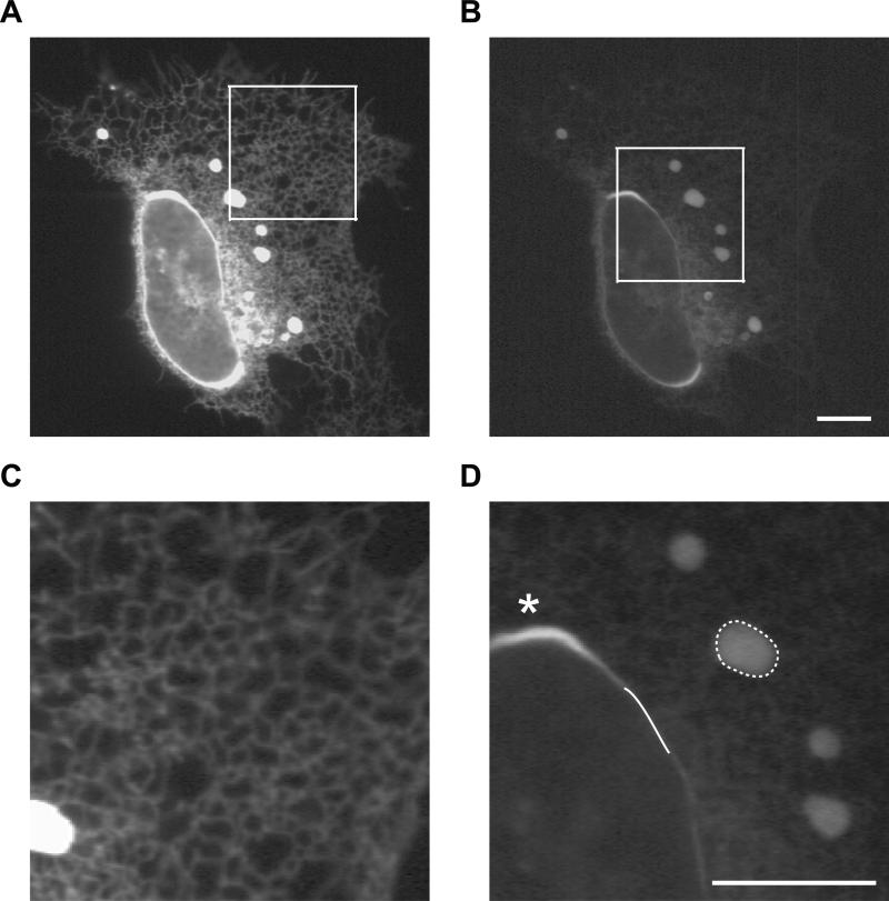
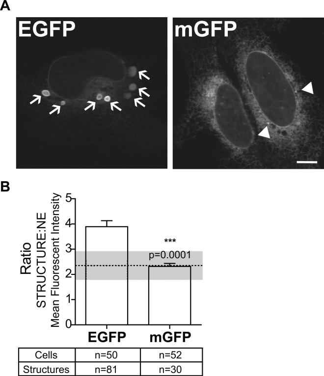
Several fluorescent proteins (FPs) are prone to forming low-affinity oligomers. This undesirable tendency is exacerbated when FPs are confined to membranes or when fused to naturally oligomeric proteins. Oligomerization of FPs limits their suitability for creating fusions with proteins of interest. Unfortunately, no standardized method evaluates the biologically relevant oligomeric state of FPs. Here, we describe a quantitative visual assay for assessing whether FPs are sufficiently monomeric under physiologic conditions. Membrane-associated FP-fusion proteins, by virtue of their constrained planar geometry, achieve high effective concentrations. We exploited this propensity to develop an assay to measure FP tendencies to oligomerize in cells. FPs were fused on the cytoplasmic end of an endoplasmic reticulum (ER) signal-anchor membrane protein (CytERM) and expressed in cells. Cells were scored based on the ability of CytERM to homo-oligomerize with proteins on apposing membranes and restructure the ER from a tubular network into organized smooth ER (OSER) whorl structures. The ratio of nuclear envelope and OSER structures mean fluorescent intensities for cells expressing enhanced green fluorescent protein (EGFP) or monomeric green fluorescent protein (mGFP) CytERM established standards for comparison of uncharacterized FPs. We tested three FPs and identified two as sufficiently monomeric, while a third previously reported as monomeric was found to strongly oligomerize.
几种荧光蛋白(FPs)易于形成低亲和力的寡聚体。当 FPs 被局限于膜内或与天然寡聚蛋白融合时,这种不理想的趋势会加剧。FPs 的寡聚化限制了它们与感兴趣的蛋白质融合的适用性。不幸的是,没有标准化的方法来评估 FPs 在生物学上相关的寡聚状态。在这里,我们描述了一种定量的可视化测定方法,用于评估 FPs 在生理条件下是否足够单体。由于其受限制的平面几何形状,膜相关的 FP-融合蛋白达到了高的有效浓度。我们利用这种倾向开发了一种测定方法,以测量 FP 在细胞中寡聚化的趋势。将 FP 融合到内质网(ER)信号锚定膜蛋白(CytERM)的细胞质末端,并在细胞中表达。根据 CytERM 与相邻膜上的蛋白质同源寡聚化的能力以及将 ER 从管状网络重构为有组织的光滑 ER(OSER)涡旋结构来对细胞进行评分。表达增强型绿色荧光蛋白(EGFP)或单体绿色荧光蛋白(mGFP)CytERM 的核膜和 OSER 结构的平均荧光强度比为未表征的 FPs 提供了比较标准。我们测试了三种 FPs,并确定了两种足够单体,而第三种先前报道为单体的 FPs 则被发现强烈寡聚化。