• 文献检索
  • 文档翻译
  • 深度研究
  • 学术资讯
  • Suppr Zotero 插件Zotero 插件
  • 邀请有礼
  • 套餐&价格
  • 历史记录
应用&插件
Suppr Zotero 插件Zotero 插件浏览器插件Mac 客户端Windows 客户端微信小程序
定价
高级版会员购买积分包购买API积分包
服务
文献检索文档翻译深度研究API 文档MCP 服务
关于我们
关于 Suppr公司介绍联系我们用户协议隐私条款
关注我们

Suppr 超能文献

核心技术专利:CN118964589B侵权必究
粤ICP备2023148730 号-1Suppr @ 2026

文献检索

告别复杂PubMed语法,用中文像聊天一样搜索,搜遍4000万医学文献。AI智能推荐,让科研检索更轻松。

立即免费搜索

文件翻译

保留排版,准确专业,支持PDF/Word/PPT等文件格式,支持 12+语言互译。

免费翻译文档

深度研究

AI帮你快速写综述,25分钟生成高质量综述,智能提取关键信息,辅助科研写作。

立即免费体验

基于 T2 加权心血管磁共振的危险心肌半自动分割。

Semi-automatic segmentation of myocardium at risk in T2-weighted cardiovascular magnetic resonance.

机构信息

Department of Clinical Physiology, Skåne University Hospital, Lund University, Lund, Sweden.

出版信息

J Cardiovasc Magn Reson. 2012 Jan 31;14(1):10. doi: 10.1186/1532-429X-14-10.

DOI:10.1186/1532-429X-14-10
PMID:22293146
原文链接:https://pmc.ncbi.nlm.nih.gov/articles/PMC3349606/
Abstract

BACKGROUND

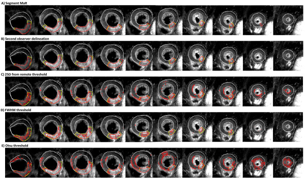
T2-weighted cardiovascular magnetic resonance (CMR) has been shown to be a promising technique for determination of ischemic myocardium, referred to as myocardium at risk (MaR), after an acute coronary event. Quantification of MaR in T2-weighted CMR has been proposed to be performed by manual delineation or the threshold methods of two standard deviations from remote (2SD), full width half maximum intensity (FWHM) or Otsu. However, manual delineation is subjective and threshold methods have inherent limitations related to threshold definition and lack of a priori information about cardiac anatomy and physiology. Therefore, the aim of this study was to develop an automatic segmentation algorithm for quantification of MaR using anatomical a priori information.

METHODS

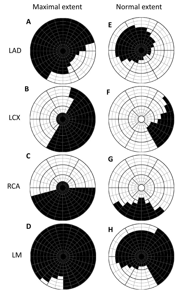
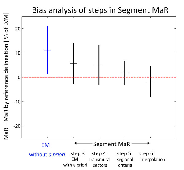
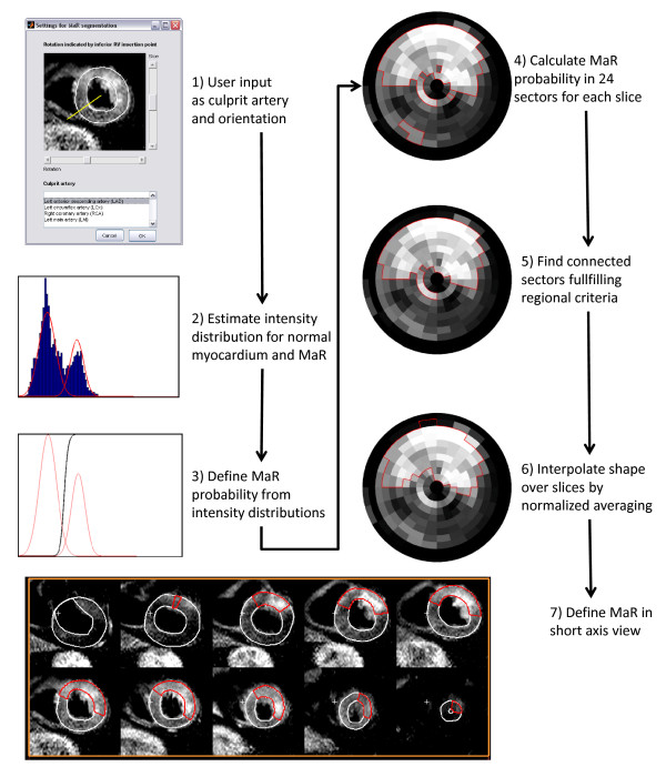
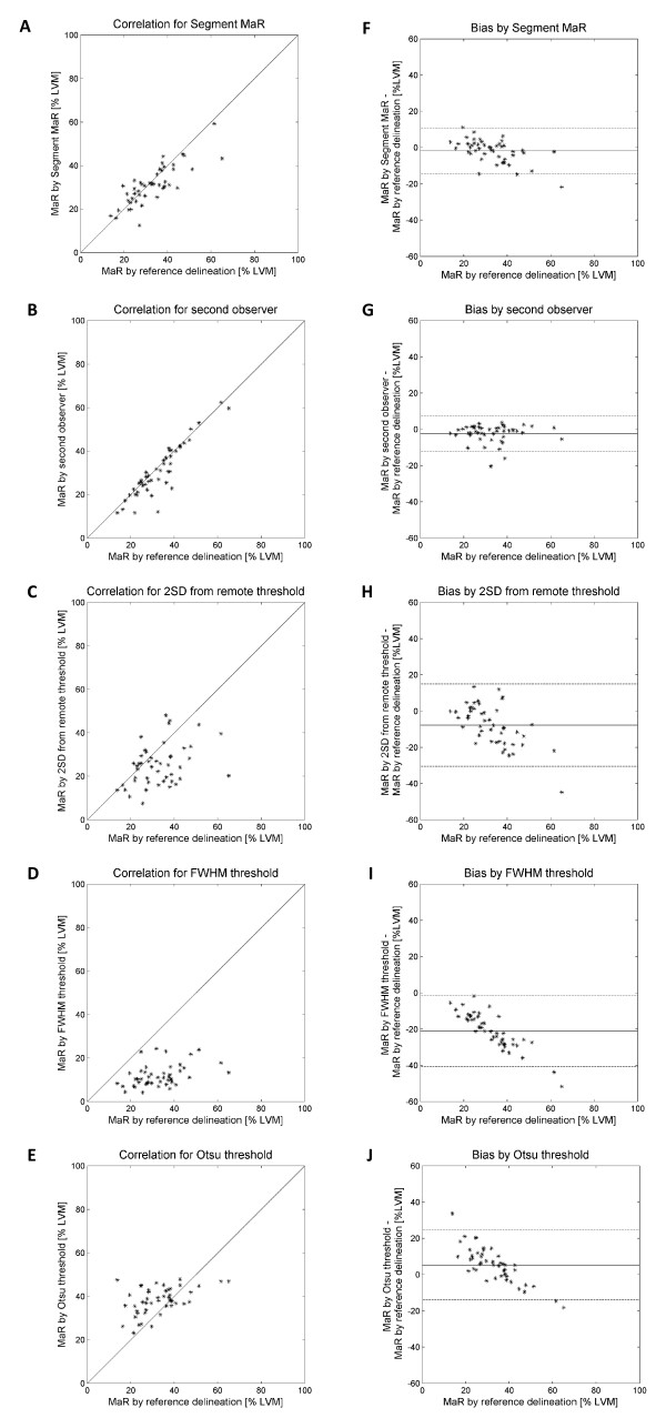
Forty-seven patients with first-time acute ST-elevation myocardial infarction underwent T2-weighted CMR within 1 week after admission. Endocardial and epicardial borders of the left ventricle, as well as the hyper enhanced MaR regions were manually delineated by experienced observers and used as reference method. A new automatic segmentation algorithm, called Segment MaR, defines the MaR region as the continuous region most probable of being MaR, by estimating the intensities of normal myocardium and MaR with an expectation maximization algorithm and restricting the MaR region by an a priori model of the maximal extent for the user defined culprit artery. The segmentation by Segment MaR was compared against inter observer variability of manual delineation and the threshold methods of 2SD, FWHM and Otsu.

RESULTS

MaR was 32.9 ± 10.9% of left ventricular mass (LVM) when assessed by the reference observer and 31.0 ± 8.8% of LVM assessed by Segment MaR. The bias and correlation was, -1.9 ± 6.4% of LVM, R = 0.81 (p < 0.001) for Segment MaR, -2.3 ± 4.9%, R = 0.91 (p < 0.001) for inter observer variability of manual delineation, -7.7 ± 11.4%, R = 0.38 (p = 0.008) for 2SD, -21.0 ± 9.9%, R = 0.41 (p = 0.004) for FWHM, and 5.3 ± 9.6%, R = 0.47 (p < 0.001) for Otsu.

CONCLUSIONS

There is a good agreement between automatic Segment MaR and manually assessed MaR in T2-weighted CMR. Thus, the proposed algorithm seems to be a promising, objective method for standardized MaR quantification in T2-weighted CMR.

摘要

背景

T2 加权心血管磁共振(CMR)已被证明是一种很有前途的技术,可用于确定急性冠状动脉事件后的缺血性心肌,即风险心肌(MaR)。T2 加权 CMR 中 MaR 的定量已被提议通过手动描绘或从远程(2SD)、全宽半最大值强度(FWHM)或 Otsu 的两个标准差的阈值方法进行。然而,手动描绘是主观的,而阈值方法具有与阈值定义相关的固有局限性,并且缺乏有关心脏解剖结构和生理学的先验信息。因此,本研究的目的是开发一种基于解剖学先验信息的自动分割算法,用于定量 MaR。

方法

47 例首次急性 ST 段抬高型心肌梗死患者在入院后 1 周内行 T2 加权 CMR。左心室的心内膜和心外膜边界以及高强化 MaR 区域由经验丰富的观察者手动描绘,并作为参考方法使用。一种新的自动分割算法,称为 Segment MaR,通过估计正常心肌和 MaR 的强度,并通过用户定义的罪魁祸首动脉的最大范围的先验模型来限制 MaR 区域,将 MaR 区域定义为最有可能是 MaR 的连续区域。分割由 Segment MaR 与手动描绘的观察者间变异性和 2SD、FWHM 和 Otsu 的阈值方法进行比较。

结果

参考观察者评估的 MaR 为左心室质量(LVM)的 32.9±10.9%,Segment MaR 评估的 MaR 为 31.0±8.8%LVM。Segment MaR 的偏倚和相关性为,-1.9±6.4%LVM,R=0.81(p<0.001);手动描绘的观察者间变异性为,-2.3±4.9%,R=0.91(p<0.001);2SD 为,-7.7±11.4%,R=0.38(p=0.008);FWHM 为,-21.0±9.9%,R=0.41(p=0.004);Otsu 为,5.3±9.6%,R=0.47(p<0.001)。

结论

在 T2 加权 CMR 中,自动 Segment MaR 与手动评估的 MaR 之间具有良好的一致性。因此,该算法似乎是一种很有前途的、客观的 T2 加权 CMR 中标准化 MaR 定量方法。

https://cdn.ncbi.nlm.nih.gov/pmc/blobs/8e2b/3349606/67ba4c471f02/1532-429X-14-10-5.jpg
https://cdn.ncbi.nlm.nih.gov/pmc/blobs/8e2b/3349606/911ab1bc91d4/1532-429X-14-10-1.jpg
https://cdn.ncbi.nlm.nih.gov/pmc/blobs/8e2b/3349606/771c3f76a6f5/1532-429X-14-10-2.jpg
https://cdn.ncbi.nlm.nih.gov/pmc/blobs/8e2b/3349606/25376bad50be/1532-429X-14-10-3.jpg
https://cdn.ncbi.nlm.nih.gov/pmc/blobs/8e2b/3349606/0e5ed54743ea/1532-429X-14-10-4.jpg
https://cdn.ncbi.nlm.nih.gov/pmc/blobs/8e2b/3349606/67ba4c471f02/1532-429X-14-10-5.jpg

相似文献

1
Semi-automatic segmentation of myocardium at risk in T2-weighted cardiovascular magnetic resonance.基于 T2 加权心血管磁共振的危险心肌半自动分割。
J Cardiovasc Magn Reson. 2012 Jan 31;14(1):10. doi: 10.1186/1532-429X-14-10.
2
Automatic segmentation of myocardium at risk from contrast enhanced SSFP CMR: validation against expert readers and SPECT.基于对比增强稳态自由进动心脏磁共振成像自动分割危险心肌:与专家阅片及单光子发射计算机断层扫描的对比验证
BMC Med Imaging. 2016 Mar 5;16:19. doi: 10.1186/s12880-016-0124-1.
3
Cardiovascular magnetic resonance of the myocardium at risk in acute reperfused myocardial infarction: comparison of T2-weighted imaging versus the circumferential endocardial extent of late gadolinium enhancement with transmural projection.急性再灌注心肌梗死心肌危险区的心血管磁共振:T2 加权成像与钆延迟增强的环形心内膜范围及透壁投影的比较。
J Cardiovasc Magn Reson. 2010 Mar 29;12(1):18. doi: 10.1186/1532-429X-12-18.
4
Highly automatic quantification of myocardial oedema in patients with acute myocardial infarction using bright blood T2-weighted CMR.应用亮血 T2 加权 CMR 对急性心肌梗死患者心肌水肿进行高度自动量化。
J Cardiovasc Magn Reson. 2013 Mar 30;15(1):28. doi: 10.1186/1532-429X-15-28.
5
Quantification of infarct size and myocardium at risk: evaluation of different techniques and its implications.梗死面积和危险心肌的定量分析:不同技术的评估及其意义
Eur Heart J Cardiovasc Imaging. 2015 Jul;16(7):738-46. doi: 10.1093/ehjci/jev001. Epub 2015 Mar 2.
6
T1 mapping and T2 mapping at 3T for quantifying the area-at-risk in reperfused STEMI patients.在3T场强下进行T1映射和T2映射以量化再灌注ST段抬高型心肌梗死患者的危险区域。
J Cardiovasc Magn Reson. 2015 Aug 12;17(1):73. doi: 10.1186/s12968-015-0173-6.
7
A new automatic algorithm for quantification of myocardial infarction imaged by late gadolinium enhancement cardiovascular magnetic resonance: experimental validation and comparison to expert delineations in multi-center, multi-vendor patient data.一种用于定量评估延迟钆增强心血管磁共振成像心肌梗死的新型自动算法:多中心、多设备患者数据的实验验证及与专家划定结果的比较
J Cardiovasc Magn Reson. 2016 May 4;18(1):27. doi: 10.1186/s12968-016-0242-5.
8
Experimental validation of contrast-enhanced SSFP cine CMR for quantification of myocardium at risk in acute myocardial infarction.对比增强稳态自由进动电影磁共振成像用于急性心肌梗死中危险心肌定量的实验验证
J Cardiovasc Magn Reson. 2017 Jan 30;19(1):12. doi: 10.1186/s12968-017-0325-y.
9
Assessment of myocardium at risk with contrast enhanced steady-state free precession cine cardiovascular magnetic resonance compared to single-photon emission computed tomography.对比增强稳态自由进动电影心血管磁共振与单光子发射计算机断层扫描评估心肌风险。
J Cardiovasc Magn Reson. 2010 Apr 30;12(1):25. doi: 10.1186/1532-429X-12-25.
10
Sources of variability in quantification of cardiovascular magnetic resonance infarct size - reproducibility among three core laboratories.心血管磁共振梗死面积定量的变异性来源 - 三个核心实验室之间的可重复性。
J Cardiovasc Magn Reson. 2017 Aug 11;19(1):62. doi: 10.1186/s12968-017-0378-y.

引用本文的文献

1
Myocardial strain analysis by feature tracking cardiac magnetic resonance to identify subclinical cardiac dysfunction in patients with MINOCA.采用特征追踪心脏磁共振分析心肌应变,以识别 MINOCA 患者的亚临床心功能障碍。
J Cardiothorac Surg. 2024 Oct 10;19(1):602. doi: 10.1186/s13019-024-03093-z.
2
Validation of contrast enhanced cine steady-state free precession and T2-weighted CMR for assessment of ischemic myocardial area-at-risk in the presence of reperfusion injury.在存在再灌注损伤的情况下,对比增强电影稳态自由进动和T2加权心脏磁共振成像用于评估缺血心肌危险区域的验证。
Int J Cardiovasc Imaging. 2019 Jun;35(6):1039-1045. doi: 10.1007/s10554-019-01569-x. Epub 2019 Mar 9.
3

本文引用的文献

1
Assessment of tissue edema in patients with acute myocardial infarction by computer-assisted quantification of triple inversion recovery prepared MRI of the myocardium.采用计算机辅助定量分析心肌三反转恢复准备 MRI 评估急性心肌梗死患者的组织水肿。
Magn Reson Med. 2011 Aug;66(2):564-73. doi: 10.1002/mrm.22812. Epub 2011 Mar 9.
2
Design and validation of Segment--freely available software for cardiovascular image analysis.设计和验证——用于心血管图像分析的免费软件段。
BMC Med Imaging. 2010 Jan 11;10:1. doi: 10.1186/1471-2342-10-1.
3
Intramyocardial hemorrhage and microvascular obstruction after primary percutaneous coronary intervention.
Infarct size following complete revascularization in patients presenting with STEMI: a comparison of immediate and staged in-hospital non-infarct related artery PCI subgroups in the CvLPRIT study.
ST段抬高型心肌梗死患者完全血运重建后的梗死面积:CvLPRIT研究中直接与分期住院非梗死相关动脉PCI亚组的比较
J Cardiovasc Magn Reson. 2016 Nov 9;18(1):85. doi: 10.1186/s12968-016-0298-2.
4
A new automatic algorithm for quantification of myocardial infarction imaged by late gadolinium enhancement cardiovascular magnetic resonance: experimental validation and comparison to expert delineations in multi-center, multi-vendor patient data.一种用于定量评估延迟钆增强心血管磁共振成像心肌梗死的新型自动算法:多中心、多设备患者数据的实验验证及与专家划定结果的比较
J Cardiovasc Magn Reson. 2016 May 4;18(1):27. doi: 10.1186/s12968-016-0242-5.
5
Cardiovascular MR T2-STIR imaging does not discriminate between intramyocardial haemorrhage and microvascular obstruction during the subacute phase of a reperfused myocardial infarction.在再灌注心肌梗死的亚急性期,心血管磁共振T2加权短tau反转恢复成像无法区分心肌内出血和微血管阻塞。
Open Heart. 2016 Apr 20;3(1):e000346. doi: 10.1136/openhrt-2015-000346. eCollection 2016.
6
Multi-vendor, multicentre comparison of contrast-enhanced SSFP and T2-STIR CMR for determining myocardium at risk in ST-elevation myocardial infarction.多厂商、多中心对比增强稳态自由进动序列(SSFP)和T2加权短反转恢复序列(T2-STIR)心脏磁共振成像用于确定ST段抬高型心肌梗死中危险心肌的比较研究
Eur Heart J Cardiovasc Imaging. 2016 Jul;17(7):744-53. doi: 10.1093/ehjci/jew027. Epub 2016 Mar 21.
7
Automatic segmentation of myocardium at risk from contrast enhanced SSFP CMR: validation against expert readers and SPECT.基于对比增强稳态自由进动心脏磁共振成像自动分割危险心肌:与专家阅片及单光子发射计算机断层扫描的对比验证
BMC Med Imaging. 2016 Mar 5;16:19. doi: 10.1186/s12880-016-0124-1.
8
Complete Versus Lesion-Only Primary PCI: The Randomized Cardiovascular MR CvLPRIT Substudy.完全血运重建与仅针对病变的直接经皮冠状动脉介入治疗:随机心血管磁共振CvLPRIT子研究
J Am Coll Cardiol. 2015 Dec 22;66(24):2713-2724. doi: 10.1016/j.jacc.2015.09.099.
9
Comparison of semi-automated methods to quantify infarct size and area at risk by cardiovascular magnetic resonance imaging at 1.5T and 3.0T field strengths.在1.5T和3.0T场强下,通过心血管磁共振成像对半自动方法定量梗死面积和危险区域的比较。
BMC Res Notes. 2015 Feb 25;8:52. doi: 10.1186/s13104-015-1007-1.
10
Quantification of infarct size and myocardium at risk: evaluation of different techniques and its implications.梗死面积和危险心肌的定量分析:不同技术的评估及其意义
Eur Heart J Cardiovasc Imaging. 2015 Jul;16(7):738-46. doi: 10.1093/ehjci/jev001. Epub 2015 Mar 2.
心肌内出血和经皮冠状动脉介入治疗后的微血管阻塞。
Int J Cardiovasc Imaging. 2010 Jan;26(1):49-55. doi: 10.1007/s10554-009-9499-1. Epub 2009 Sep 15.
4
Quantification of myocardial area at risk with T2-weighted CMR: comparison with contrast-enhanced CMR and coronary angiography.采用T2加权心脏磁共振成像定量评估心肌梗死危险区:与对比增强心脏磁共振成像及冠状动脉造影的比较
JACC Cardiovasc Imaging. 2009 Jul;2(7):825-31. doi: 10.1016/j.jcmg.2009.02.011.
5
Myocardium at risk after acute infarction in humans on cardiac magnetic resonance: quantitative assessment during follow-up and validation with single-photon emission computed tomography.人类急性心肌梗死后心脏磁共振成像显示的危险心肌:随访期间的定量评估及与单光子发射计算机断层扫描的验证
JACC Cardiovasc Imaging. 2009 May;2(5):569-76. doi: 10.1016/j.jcmg.2008.11.018.
6
The salvaged area at risk in reperfused acute myocardial infarction as visualized by cardiovascular magnetic resonance.通过心血管磁共振成像观察到的再灌注急性心肌梗死中存在风险的挽救区域。
J Am Coll Cardiol. 2008 Apr 22;51(16):1581-7. doi: 10.1016/j.jacc.2008.01.019.
7
Morphometric analysis of white matter lesions in MR images: method and validation.磁共振图像中脑白质病变的形态计量分析:方法与验证。
IEEE Trans Med Imaging. 1994;13(4):716-24. doi: 10.1109/42.363096.
8
In vivo T2-weighted magnetic resonance imaging can accurately determine the ischemic area at risk for 2-day-old nonreperfused myocardial infarction.活体T2加权磁共振成像能够准确测定2日龄未再灌注心肌梗死的危险缺血区域。
Invest Radiol. 2008 Jan;43(1):7-15. doi: 10.1097/RLI.0b013e3181558822.
9
T2-weighted cardiovascular magnetic resonance imaging.T2加权心血管磁共振成像。
J Magn Reson Imaging. 2007 Sep;26(3):452-9. doi: 10.1002/jmri.21028.
10
Value of T2-weighted, first-pass and delayed enhancement, and cine CMR to differentiate between acute and chronic myocardial infarction.T2加权成像、首过灌注及延迟强化成像以及心脏磁共振电影成像在鉴别急性和慢性心肌梗死中的价值。
Eur Radiol. 2007 Mar;17(3):610-7. doi: 10.1007/s00330-006-0460-6. Epub 2006 Dec 6.