Psychopharmacology Section, Division of Psychiatry, University of Nottingham, Room B109, Medical School, Queen's Medical Centre, Nottingham NG7 2UH, UK.
Behav Brain Res. 2012 Apr 15;229(2):372-7. doi: 10.1016/j.bbr.2012.01.035. Epub 2012 Jan 24.
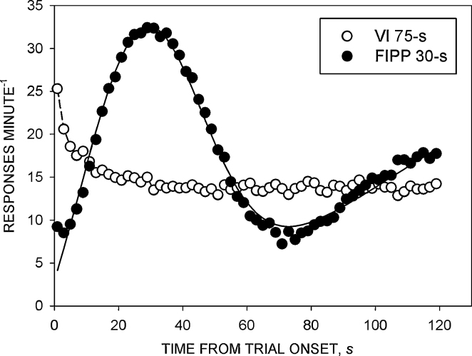
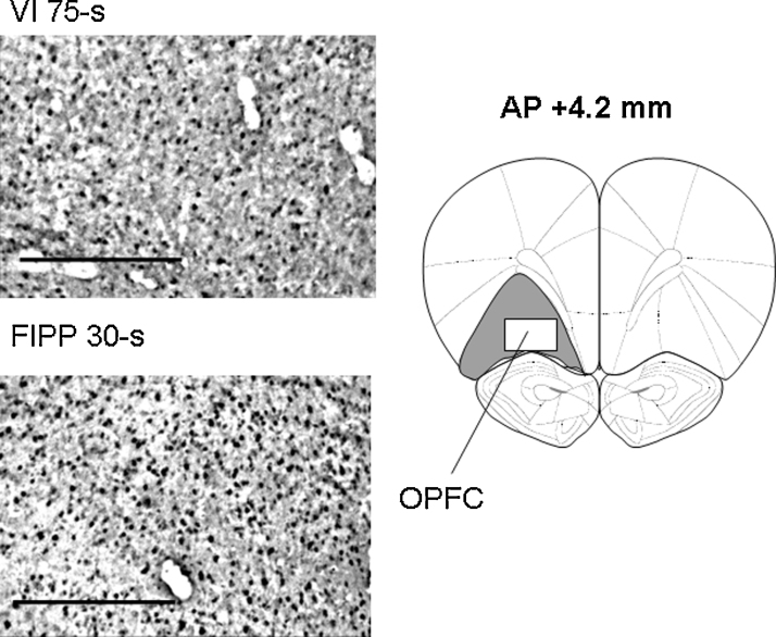
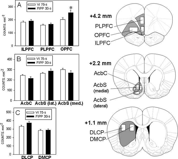
It has been proposed that cortico-striato-thalamo-cortical circuits that incorporate the prefrontal cortex and dorsal striatum regulate interval timing behaviour. The present experiment examined whether performance on the fixed-interval peak procedure (FIPP), an immediate timing schedule, would induce neuronal activity in cortical and striatal areas, as revealed by enhanced expression of the Fos protein, a marker for neuronal activation. Regional Fos expression was compared between rats trained on the FIPP and rats trained on a variable-interval (VI) schedule matched to the FIPP for overall response rate and reinforcer delivery. Response rate in the peak trials of the FIPP conformed to a temporally differentiated pattern, which was well described by a modified Gaussian function; in agreement with previous findings, the peak time occurred close to the time at which the reinforcer was delivered in the fixed-interval trials, and the Weber fraction was within the range of values reported previously. The density of Fos-positive neurones (counts mm(-2)) in the orbital prefrontal cortex (OPFC) was greater in rats exposed to the FIPP than in rats exposed to the VI schedule, suggesting a greater activation of this area during the performance of the former task. This is consistent with the results of previous studies that have implicated the OPFC in interval timing behaviour. However, there was no significant difference between the levels of Fos expression in the dorsal or ventral striatum of the rats trained under the two schedules.
有人提出,包含前额皮质和背侧纹状体的皮质-纹状体-丘脑-皮质回路调节间隔时间行为。本实验研究了在固定间隔峰值程序(FIPP)上的表现是否会诱导皮质和纹状体区域的神经元活动,如 Fos 蛋白的增强表达所揭示的,Fos 蛋白是神经元激活的标志物。将在 FIPP 上接受训练的大鼠和在与 FIPP 相匹配的可变间隔(VI)方案上接受训练的大鼠的区域 Fos 表达进行了比较,以达到整体反应率和强化物传递的匹配。FIPP 中的峰值试验的反应率符合时间分化模式,该模式由改进的高斯函数很好地描述;与先前的发现一致,峰值时间接近在固定间隔试验中传递强化物的时间,Weber 分数在先前报告的范围内。暴露于 FIPP 的大鼠的眶额前皮质(OPFC)中的 Fos 阳性神经元(计数 mm(-2))密度高于暴露于 VI 方案的大鼠,这表明在执行前者任务期间该区域的激活更大。这与先前的研究结果一致,这些研究表明 OPFC 参与了间隔时间行为。然而,在两种方案下接受训练的大鼠的背侧或腹侧纹状体中的 Fos 表达水平没有显著差异。