• 文献检索
  • 文档翻译
  • 深度研究
  • 学术资讯
  • Suppr Zotero 插件Zotero 插件
  • 邀请有礼
  • 套餐&价格
  • 历史记录
应用&插件
Suppr Zotero 插件Zotero 插件浏览器插件Mac 客户端Windows 客户端微信小程序
定价
高级版会员购买积分包购买API积分包
服务
文献检索文档翻译深度研究API 文档MCP 服务
关于我们
关于 Suppr公司介绍联系我们用户协议隐私条款
关注我们

Suppr 超能文献

核心技术专利:CN118964589B侵权必究
粤ICP备2023148730 号-1Suppr @ 2026

文献检索

告别复杂PubMed语法,用中文像聊天一样搜索,搜遍4000万医学文献。AI智能推荐,让科研检索更轻松。

立即免费搜索

文件翻译

保留排版,准确专业,支持PDF/Word/PPT等文件格式,支持 12+语言互译。

免费翻译文档

深度研究

AI帮你快速写综述,25分钟生成高质量综述,智能提取关键信息,辅助科研写作。

立即免费体验

一项关于在进行三维(3D)头影测量分析时,头影测量标志点的可重复性的研究。

A study on the reproducibility of cephalometric landmarks when undertaking a three-dimensional (3D) cephalometric analysis.

机构信息

Department of Orthodontics, Faculty of Medicine and Dentistry, University of Valencia, Valencia, Spain.

出版信息

Med Oral Patol Oral Cir Bucal. 2012 Jul 1;17(4):e678-88. doi: 10.4317/medoral.17721.

DOI:10.4317/medoral.17721
PMID:22322503
原文链接:https://pmc.ncbi.nlm.nih.gov/articles/PMC3476034/
Abstract

OBJECTIVES

Cone Beam Computerized Tomography (CBCT) allows the possibility of modifying some of the diagnostic tools used in orthodontics, such as cephalometry. The first step must be to study the characteristics of these devices in terms of accuracy and reliability of the most commonly used landmarks. The aims were 1- To assess intra and inter-observer reliability in the location of anatomical landmarks belonging to hard tissues of the skull in images taken with a CBCT device, 2- To determine which of those landmarks are more vs. less reliable and 3- To introduce planes of reference so as to create cephalometric analyses appropriated to the 3D reality.

STUDY DESIGN

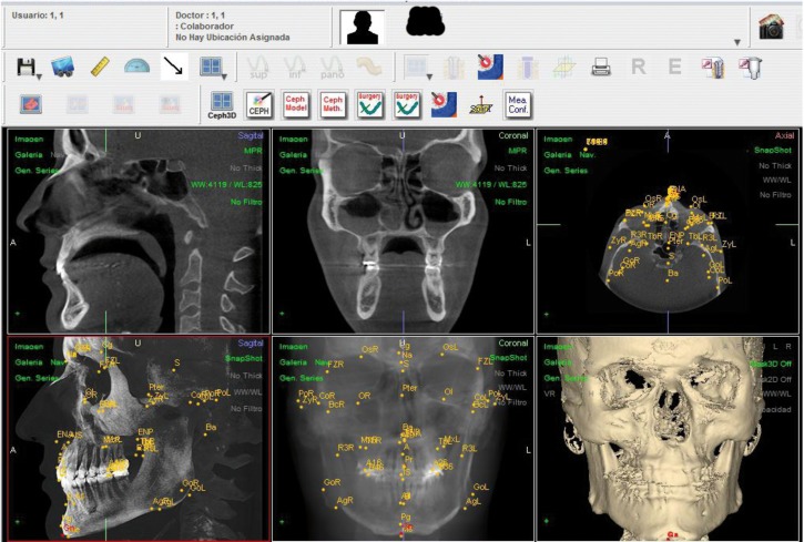
Fifteen patients who had a CBCT (i-CAT) as a diagnostic register were selected. To assess the reproducibility on landmark location and the differences in the measurements of two observers at different times, 41 landmarks were defined on the three spatial axes (X,Y,Z) and located. 3.690 measurements were taken and, as each determination has 3 coordinates, 11.070 data were processed with SPSS statistical package. To discover the reproducibility of the method on landmark location, an ANOVA was undertaken using two variation factors: time (t1, t2 and t3) and observer (Ob1 and Ob2) for each axis (X, Y and Z) and landmark. The order of the CBCT scans submitted to the observers (Ob1, Ob2) at t1, t2, and t3, were different and randomly allocated. Multiple comparisons were undertaken using the Bonferroni test. The intra- and inter-examiner ICC's were calculated.

RESULTS

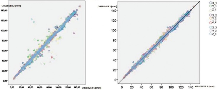
Intra- and interexaminer reliability was high, both being ICC ≥ 0.99, with the best frequency on axis Z.

CONCLUSIONS

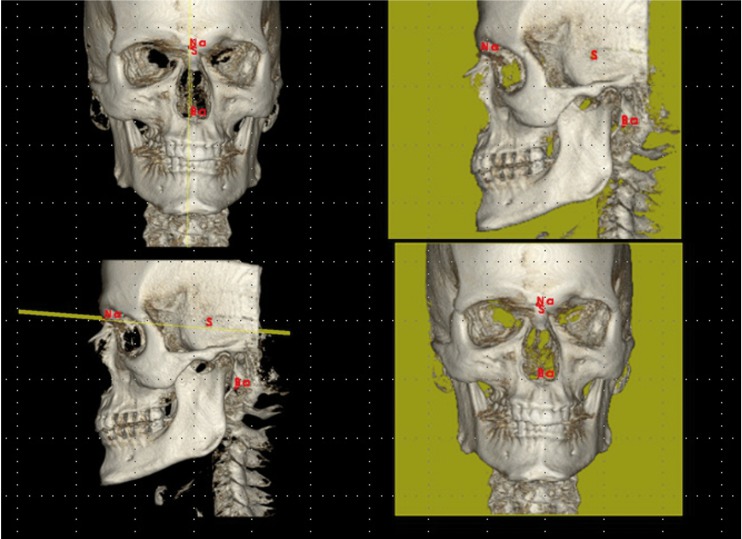
The most reliable landmarks were: Nasion, Sella, Basion, left Porion, point A, anterior nasal spine, Pogonion, Gnathion, Menton, frontozygomatic sutures, first lower molars and upper and lower incisors. Those with less reliability were the supraorbitals, right zygion and posterior nasal spine.

摘要

目的

锥形束计算机断层扫描(CBCT)使得修改一些在正畸学中使用的诊断工具成为可能,如头影测量。第一步必须研究这些设备在最常用的解剖标志的准确性和可靠性方面的特征。目的是:1. 评估在使用 CBCT 设备拍摄的图像中,属于颅骨硬组织的解剖标志的定位的观察者内和观察者间的可靠性,2. 确定哪些标志更可靠,哪些标志不太可靠,3. 引入参考平面,以便创建适用于 3D 现实的头影测量分析。

研究设计

选择了 15 名患者,他们使用 CBCT(i-CAT)作为诊断记录。为了评估在定位地标位置方面的可重复性和两位观察者在不同时间的测量值之间的差异,在三个空间轴(X、Y、Z)上定义了 41 个地标并进行了定位。共进行了 3.690 次测量,由于每次测定有 3 个坐标,共处理了 11.070 个数据,使用 SPSS 统计软件包进行处理。为了发现地标定位方法的可重复性,采用方差分析方法,使用两个变化因素:时间(t1、t2 和 t3)和观察者(Ob1 和 Ob2),用于每个轴(X、Y 和 Z)和地标。观察者(Ob1、Ob2)在 t1、t2 和 t3 时提交给观察者的 CBCT 扫描的顺序不同,并且是随机分配的。使用 Bonferroni 检验进行多重比较。计算了观察者内和观察者间的 ICC。

结果

观察者内和观察者间的可靠性都很高,ICC 均≥0.99,Z 轴上的频率最高。

结论

最可靠的地标是:Nasion、Sella、Basion、左侧 Porion、点 A、前鼻棘、Pogonion、Gnathion、Menton、额颧缝、第一下磨牙以及上下切牙。可靠性较低的地标是眶上嵴、右侧颧突和后鼻棘。

https://cdn.ncbi.nlm.nih.gov/pmc/blobs/b634/3476034/1c9ee9b09c97/medoral-17-e678-g003.jpg
https://cdn.ncbi.nlm.nih.gov/pmc/blobs/b634/3476034/6675f25a96eb/medoral-17-e678-g001.jpg
https://cdn.ncbi.nlm.nih.gov/pmc/blobs/b634/3476034/264daf121505/medoral-17-e678-g002.jpg
https://cdn.ncbi.nlm.nih.gov/pmc/blobs/b634/3476034/1c9ee9b09c97/medoral-17-e678-g003.jpg
https://cdn.ncbi.nlm.nih.gov/pmc/blobs/b634/3476034/6675f25a96eb/medoral-17-e678-g001.jpg
https://cdn.ncbi.nlm.nih.gov/pmc/blobs/b634/3476034/264daf121505/medoral-17-e678-g002.jpg
https://cdn.ncbi.nlm.nih.gov/pmc/blobs/b634/3476034/1c9ee9b09c97/medoral-17-e678-g003.jpg

相似文献

1
A study on the reproducibility of cephalometric landmarks when undertaking a three-dimensional (3D) cephalometric analysis.一项关于在进行三维(3D)头影测量分析时,头影测量标志点的可重复性的研究。
Med Oral Patol Oral Cir Bucal. 2012 Jul 1;17(4):e678-88. doi: 10.4317/medoral.17721.
2
Reliability of anatomic structures as landmarks in three-dimensional cephalometric analysis using CBCT.使用锥形束计算机断层扫描(CBCT)进行三维头影测量分析时,解剖结构作为标志点的可靠性。
Angle Orthod. 2014 Sep;84(5):762-72. doi: 10.2319/090413-652.1. Epub 2013 Dec 23.
3
Comparison of observer reliability of three-dimensional cephalometric landmark identification on subject images from Galileos and i-CAT cone beam CT.比较 Galileos 和 i-CAT 锥形束 CT 受试者图像上三维头影测量标志点识别的观察者可靠性。
Dentomaxillofac Radiol. 2013;42(9):20130059. doi: 10.1259/dmfr.20130059. Epub 2013 Jul 5.
4
Reliability of cephalometric landmark identification on three-dimensional computed tomographic images.三维计算机断层扫描图像上头影测量标志点识别的可靠性
Br J Oral Maxillofac Surg. 2022 Apr;60(3):320-325. doi: 10.1016/j.bjoms.2021.07.003. Epub 2021 Jul 17.
5
A new mandible-specific landmark reference system for three-dimensional cephalometry using cone-beam computed tomography.一种使用锥形束计算机断层扫描的用于三维头影测量的新型下颌骨特定标志点参考系统。
Eur J Orthod. 2016 Dec;38(6):563-568. doi: 10.1093/ejo/cjv088. Epub 2015 Dec 17.
6
Intraexaminer and interexaminer reliabilities of landmark identification on digitized lateral cephalograms and formatted 3-dimensional cone-beam computerized tomography images.数字化侧位头颅影像和格式化三维锥形束计算机断层扫描影像上标志点识别的观察者内和观察者间可靠性。
Am J Orthod Dentofacial Orthop. 2010 May;137(5):598-604. doi: 10.1016/j.ajodo.2008.07.018.
7
Comparison of reliability in anatomical landmark identification using two-dimensional digital cephalometrics and three-dimensional cone beam computed tomography in vivo.在体使用二维数字头影测量法和三维锥形束计算机断层扫描术进行解剖标志点识别的可靠性比较。
Dentomaxillofac Radiol. 2009 Jul;38(5):262-73. doi: 10.1259/dmfr/81889955.
8
3D landmarks of Craniofacial Imaging and subsequent considerations on superimpositions in orthodontics-The Aarhus perspective.颅面成像的 3D 标志点及其在正畸学中的叠加后续考虑——奥胡斯视角。
Orthod Craniofac Res. 2019 May;22 Suppl 1:21-29. doi: 10.1111/ocr.12299.
9
Evaluation of the accuracy of linear measurements on lateral cephalograms obtained from cone-beam computed tomography scans with digital lateral cephalometric radiography: an in vitro study.锥束计算机断层扫描与数字化侧位头影测量X线摄影所得头颅侧位片上线性测量准确性的评估:一项体外研究
J Craniofac Surg. 2014 Sep;25(5):1710-3. doi: 10.1097/SCS.0000000000000908.
10
In vivo reliability of 3D cephalometric landmark determination on magnetic resonance imaging: a feasibility study.磁共振成像中三维头影测量标志点确定的体内可靠性:一项可行性研究。
Clin Oral Investig. 2020 Mar;24(3):1339-1349. doi: 10.1007/s00784-019-03015-7. Epub 2019 Jul 27.

引用本文的文献

1
Cranial Vault Deformation and Its Association with Mandibular Deviation in Patients with Facial Asymmetry: A CT-Based Study.颅骨穹窿变形及其与面部不对称患者下颌偏斜的关联:一项基于CT的研究。
Diagnostics (Basel). 2025 Jul 3;15(13):1702. doi: 10.3390/diagnostics15131702.
2
Evaluating the precision and reproducibility of cephalometric landmarks in locally reconstructed lateral cephalometric radiographs from cone beam computed tomography (CBCT).评估锥形束计算机断层扫描(CBCT)局部重建的头颅侧位X线片上头影测量标志点的精度和可重复性。
BMC Oral Health. 2025 May 31;25(1):866. doi: 10.1186/s12903-025-06258-x.
3
Artificial intelligence-based predictive model for guidance on treatment strategy selection in oral and maxillofacial surgery.

本文引用的文献

1
Consistency and precision of landmark identification in three-dimensional cone beam computed tomography scans.三维锥形束计算机断层扫描中标志点识别的一致性和精确性。
Eur J Orthod. 2012 Jun;34(3):263-75. doi: 10.1093/ejo/cjq144. Epub 2011 Mar 8.
2
Influence of a programme of professional calibration in the variability of landmark identification using cone beam computed tomography-synthesized and conventional radiographic cephalograms.应用锥形束 CT 合成头影测量与传统头影测量技术对标志点识别的可重复性的影响:专业标定方案的作用
Dentomaxillofac Radiol. 2010 Oct;39(7):414-23. doi: 10.1259/dmfr/31924248.
3
Intraexaminer and interexaminer reliabilities of landmark identification on digitized lateral cephalograms and formatted 3-dimensional cone-beam computerized tomography images.
基于人工智能的口腔颌面外科治疗策略选择指导预测模型。
Heliyon. 2024 Aug 2;10(15):e35742. doi: 10.1016/j.heliyon.2024.e35742. eCollection 2024 Aug 15.
4
Utilization of a Cortical Xenogeneic Membrane for Guided Bone Regeneration: A Retrospective Case Series.利用皮质异种膜进行引导性骨再生:一项回顾性病例系列研究。
J Clin Med. 2024 Aug 5;13(15):4575. doi: 10.3390/jcm13154575.
5
Skeletal facial asymmetry: reliability of manual and artificial intelligence-driven analysis.骨骼面部不对称:手动和人工智能驱动分析的可靠性。
Dentomaxillofac Radiol. 2024 Jan 11;53(1):52-59. doi: 10.1093/dmfr/twad006.
6
Three-dimensional quantification of mandibular asymmetries in Caucasian adult patients with different sagittal and vertical skeletal patterns. A cone beam study using 3D segmentation and mirroring procedures.不同矢状面和垂直骨面型的白种成人患者下颌不对称的三维定量研究。使用 3D 分割和镜像程序的锥形束研究。
Head Face Med. 2023 Dec 14;19(1):54. doi: 10.1186/s13005-023-00400-2.
7
The Association Between Skeletal Facial Types and Third Molars Impaction in a Saudi Arabian Subpopulation: A CBCT Study.沙特阿拉伯亚人群中骨骼面部类型与第三磨牙阻生之间的关联:一项CBCT研究
Clin Cosmet Investig Dent. 2023 Aug 15;15:143-156. doi: 10.2147/CCIDE.S419325. eCollection 2023.
8
Automated detection of cephalometric landmarks using deep neural patchworks.基于深度神经网络补丁的头影测量标志点自动检测
Dentomaxillofac Radiol. 2023 Sep;52(6):20230059. doi: 10.1259/dmfr.20230059. Epub 2023 Jul 3.
9
The Maxilla-Mandibular Discrepancies through Soft-Tissue References: Reliability and Validation of the Anteroposterior Measurement.通过软组织参考评估上颌-下颌差异:前后测量的可靠性与验证
Children (Basel). 2023 Feb 26;10(3):459. doi: 10.3390/children10030459.
10
Intra- and Interexaminer Measurement Variability Analysis of an Orthodontic Gauge Device to Determine Incisor Occlusal Surface Angles in the Horse.用于确定马切牙咬合面角度的正畸测量仪的检查者内和检查者间测量变异性分析。
Vet Sci. 2022 Sep 7;9(9):481. doi: 10.3390/vetsci9090481.
数字化侧位头颅影像和格式化三维锥形束计算机断层扫描影像上标志点识别的观察者内和观察者间可靠性。
Am J Orthod Dentofacial Orthop. 2010 May;137(5):598-604. doi: 10.1016/j.ajodo.2008.07.018.
4
A comparison between 2D and 3D cephalometry on CBCT scans of human skulls.二维和三维头影测量在人类颅骨 CBCT 扫描中的比较。
Int J Oral Maxillofac Surg. 2010 Feb;39(2):156-60. doi: 10.1016/j.ijom.2009.11.017. Epub 2009 Dec 30.
5
Reliability of traditional cephalometric landmarks as seen in three-dimensional analysis in maxillary expansion treatments.传统头影测量标志点在三维分析中对上颌扩弓治疗的可靠性。
Angle Orthod. 2009 Nov;79(6):1047-56. doi: 10.2319/010509-10R.1.
6
Cone-beam computerized tomography (CBCT) imaging of the oral and maxillofacial region: a systematic review of the literature.口腔颌面部锥形束计算机断层扫描(CBCT)成像:文献系统综述
Int J Oral Maxillofac Surg. 2009 Jun;38(6):609-25. doi: 10.1016/j.ijom.2009.02.028. Epub 2009 May 21.
7
Comparison of radiation dose for implant imaging using conventional spiral tomography, computed tomography, and cone-beam computed tomography.使用传统螺旋断层扫描、计算机断层扫描和锥形束计算机断层扫描进行植入物成像时的辐射剂量比较。
Oral Surg Oral Med Oral Pathol Oral Radiol Endod. 2009 Apr;107(4):559-65. doi: 10.1016/j.tripleo.2008.11.009. Epub 2009 Jan 25.
8
Linear accuracy of cone beam CT derived 3D images.锥形束CT衍生3D图像的线性精度。
Angle Orthod. 2009 Jan;79(1):150-7. doi: 10.2319/122407-599.1.
9
The use of cone-beam computed tomography in an orthodontic department in between research and daily clinic.锥形束计算机断层扫描在正畸科研究与日常临床中的应用。
World J Orthod. 2008 Fall;9(3):269-82.
10
Accuracy and repeatability of cone-beam computed tomography (CBCT) measurements used in the determination of facial indices in the laboratory setup.实验室环境下用于测定面部指数的锥形束计算机断层扫描(CBCT)测量的准确性和可重复性。
J Craniomaxillofac Surg. 2009 Jan;37(1):18-23. doi: 10.1016/j.jcms.2008.07.006. Epub 2008 Sep 23.