The Feinstein Institute for Medical Research, 350 Community Drive, Room 1239, Manhasset, NY 11030, USA.
Clin Cancer Res. 2012 Apr 1;18(7):1925-35. doi: 10.1158/1078-0432.CCR-11-2941. Epub 2012 Feb 9.
Respiratory papillomas, caused by human papillomaviruses types 6 and 11 (HPV6/11), are premalignant lesions with potential for malignant conversion. The cytokine and chemokine micromilieu of papillomas is T(H)2-like with a marked absence of IFN-γ expression. To illuminate why patients with recurrent respiratory papillomatosis (RRP) fail to effectively control their disease, we further investigated the suppressive cellular microenvironment in papillomas.
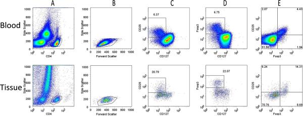
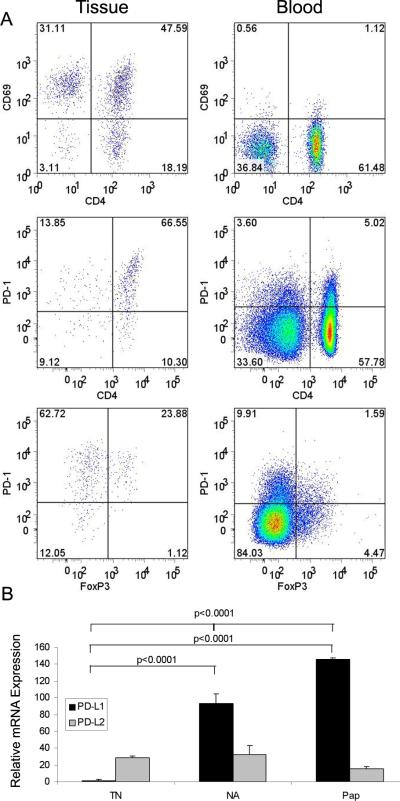
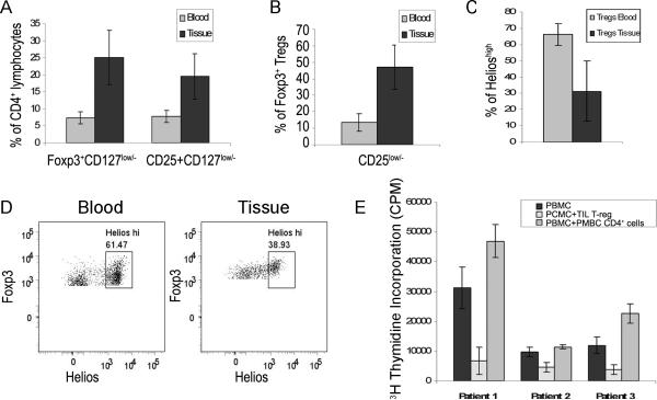
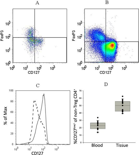
CD4(+)CD25(+)CD127(low/-)Foxp3(+) regulatory T cells (Treg) and CD4(+)CD25(-)CD127(low/-)Foxp3(-) T cells within papillomas were characterized and isolated. Their suppressor function was measured by inhibition of peripheral blood mononuclear cell (PBMC) proliferation. Expression of PD-1, CD69, and Helios was identified on these T cells. PD-L1, PD-L2, CCL17, and CCL22 mRNA was also identified in papillomas by quantitative PCR.
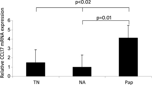
Functional Tregs were markedly enriched in papillomas and strongly inhibited anti-CD3 and anti-CD28 antibody activated PBMC proliferation. The natural Treg marker Helios was reduced on Tregs from papillomas, indicating that the majority of Tregs in papillomas are adaptive. The majority of the papilloma-derived CD4(+) T cells expressed the CD4(+)CD25(-)CD127(low/-)Foxp3(-)PD1(+)CD69(+) phenotype and failed to suppress PBMC proliferation, suggesting that they are chronically activated and exhausted. The Treg-attracting chemokine CCL22 was equally expressed by all laryngeal tissues examined. However, CCL17 was robustly expressed by papillomas compared with unaffected laryngeal tissues from RRP patients and individuals without RRP. PD-L1 was elevated in papillomas compared with control laryngeal tissues.
Papilloma CD4(+) T cells are enriched with functional Tregs, and the adaptive Helios(-) Treg fraction was increased within the T(H)2-like papilloma micromilieu. CD4(+)CD25(-)CD127(low/-)Foxp3(-) T-cells failed to suppress PBMC proliferation and may be exhausted. The PD-1/PDL-1 pathway may represent an additional immunosuppressive mechanism that contributes to defective HPV6/11 clearance in RRP.
由人乳头瘤病毒 6 型和 11 型(HPV6/11)引起的呼吸道乳头瘤是具有恶性转化潜能的癌前病变。乳头瘤的细胞因子和趋化因子微环境呈 T(H)2 样,IFN-γ 表达明显缺失。为了阐明为什么复发性呼吸道乳头瘤病(RRP)患者不能有效控制疾病,我们进一步研究了乳头瘤中的抑制性细胞微环境。
对乳头瘤中的 CD4(+)CD25(+)CD127(low/-)Foxp3(+)调节性 T 细胞(Treg)和 CD4(+)CD25(-)CD127(low/-)Foxp3(-)T 细胞进行了特征和分离。通过抑制外周血单核细胞(PBMC)增殖来测量它们的抑制功能。鉴定了这些 T 细胞上 PD-1、CD69 和 Helios 的表达。通过定量 PCR 鉴定了乳头瘤中的 PD-L1、PD-L2、CCL17 和 CCL22 mRNA。
功能性 Treg 在乳头瘤中明显富集,并强烈抑制抗 CD3 和抗 CD28 抗体激活的 PBMC 增殖。乳头瘤中的天然 Treg 标志物 Helios 减少,表明乳头瘤中的大多数 Treg 是适应性的。大多数来自乳头瘤的 CD4(+)T 细胞表达 CD4(+)CD25(-)CD127(low/-)Foxp3(-)PD1(+)CD69(+)表型,不能抑制 PBMC 增殖,表明它们处于慢性激活和衰竭状态。趋化因子 CCL22 以与所有检查的喉部组织相等的方式表达。然而,与 RRP 患者和无 RRP 个体的未受影响的喉部组织相比,CCL17 在乳头瘤中强烈表达。与对照喉部组织相比,PD-L1 在乳头瘤中升高。
乳头瘤 CD4(+)T 细胞富含功能性 Treg,在 T(H)2 样乳头瘤微环境中增加了适应性 Helios(-)Treg 分数。CD4(+)CD25(-)CD127(low/-)Foxp3(-)T 细胞不能抑制 PBMC 增殖,可能已经衰竭。PD-1/PDL-1 通路可能代表一种额外的免疫抑制机制,有助于 RRP 中 HPV6/11 的清除缺陷。