Gupta Shilpa, Kaur Mandeep, Gupta Ruchika, Singh Sompal, Pant Leela, Singh P P
Department of Pathology, Hindu Rao Hospital, Delhi, India .
Indian J Dermatol. 2011 Nov;56(6):647-9. doi: 10.4103/0019-5154.91820.
Vascular proliferation in the papillary dermis is considered to be an important and probably an early feature of psoriasis. Few morphometric studies have attempted to analyze the vascular changes. However, no study was found in the available literature comparing vascular changes between psoriasis and psoriasiform dermatitis.
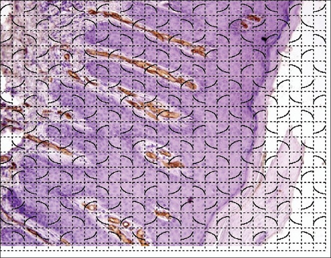
Skin biopsies from 25 cases each of psoriasis and psoriasiform lesions were immunohistochemically stained for CD34 (endothelial marker). Microvessel density (MVD), microvessel length density and ratio of microvessel area to papillary dermal area were calculated using image analysis software.
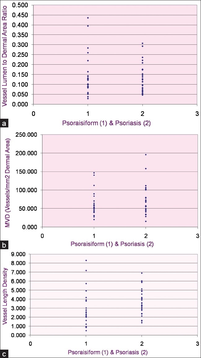
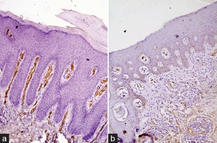
Skin biopsies from psoriasis showed higher staining for CD34 on light microscopy. Using morphometric techniques, microvessel length density was significantly higher in psoriasis compared to psoriasiform lesions (P value <0.05). MVD was also higher in psoriasis, though the difference was not significant. The ratio of microvessel area to dermal area was almost similar in both the groups.
Our results indicate that vascular tortuousity and dilatation is significant only in psoriatic lesions. These results may assist in automated diagnosis of skin biopsies.
乳头层真皮中的血管增生被认为是银屑病的一个重要且可能是早期特征。很少有形态计量学研究试图分析血管变化。然而,在现有文献中未发现比较银屑病和银屑病样皮炎之间血管变化的研究。
对25例银屑病患者和25例银屑病样皮损患者的皮肤活检组织进行CD34(内皮标志物)免疫组化染色。使用图像分析软件计算微血管密度(MVD)、微血管长度密度以及微血管面积与乳头层真皮面积的比值。
银屑病患者的皮肤活检组织在光学显微镜下显示CD34染色更强。使用形态计量学技术,银屑病患者的微血管长度密度显著高于银屑病样皮损患者(P值<0.05)。银屑病患者的MVD也较高,尽管差异不显著。两组的微血管面积与真皮面积的比值几乎相似。
我们的结果表明,血管迂曲和扩张仅在银屑病皮损中显著。这些结果可能有助于皮肤活检的自动诊断。