RIKEN Center for Developmental Biology, Kobe, Japan.
PLoS One. 2012;7(2):e31638. doi: 10.1371/journal.pone.0031638. Epub 2012 Feb 8.
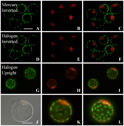
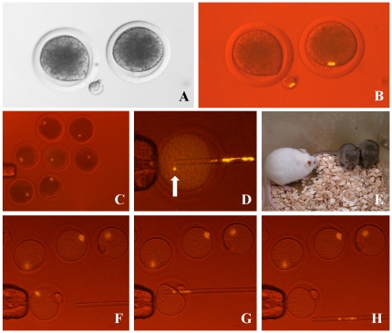
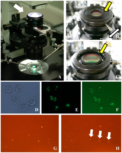
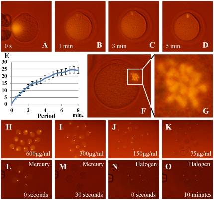
Technologies for vitally labeling cells with fluorescent dyes have advanced remarkably. However, to excite fluorescent dyes currently requires powerful illumination, which can cause phototoxic damage to the cells and increases the cost of microscopy. We have developed a filter system to excite fluorescent dyes using a conventional transmission microscope equipped with a halogen lamp. This method allows us to observe previously invisible cell organelles, such as the metaphase spindle of oocytes, without causing phototoxicity. Cells remain healthy even after intensive manipulation under fluorescence observation, such as during bovine, porcine and mouse somatic cell cloning using nuclear transfer. This method does not require expensive epifluorescence equipment and so could help to reduce the science gap between developed and developing countries.
用荧光染料对细胞进行活力标记的技术已经取得了显著的进展。然而,目前激发荧光染料需要强大的照明,这会对细胞造成光毒性损伤,并增加显微镜的成本。我们开发了一种滤光系统,可利用配备卤素灯的传统透射显微镜来激发荧光染料。这种方法使我们能够观察以前看不见的细胞细胞器,如卵母细胞的中期纺锤体,而不会造成光毒性。即使在荧光观察下进行密集的操作,例如使用核移植进行牛、猪和鼠体细胞克隆时,细胞仍然保持健康。这种方法不需要昂贵的落射荧光设备,因此有助于缩小发达国家和发展中国家之间的科学差距。