Juanpere Sergi, Perez Elsa, Huc Oscar, Motos Naiara, Pont Josep, Pedraza Salvador
Insights Imaging. 2011 Dec;2(6):653-670. doi: 10.1007/s13244-011-0122-3. Epub 2011 Aug 7.
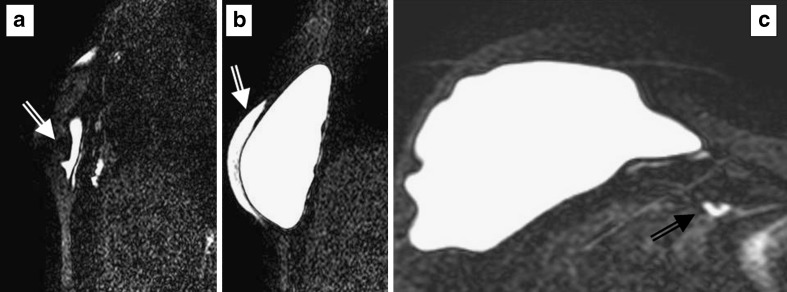
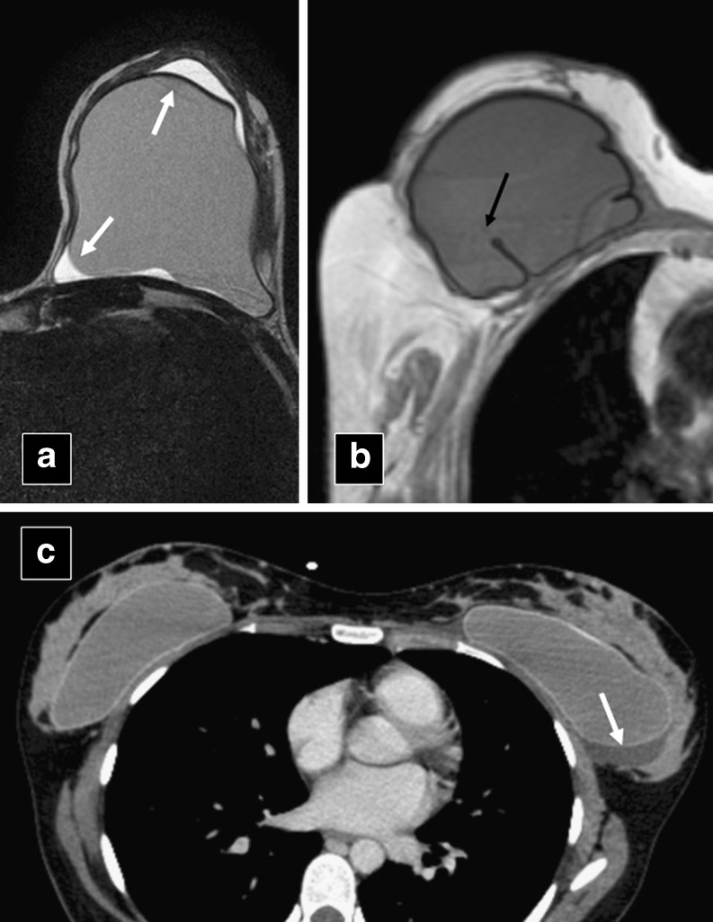
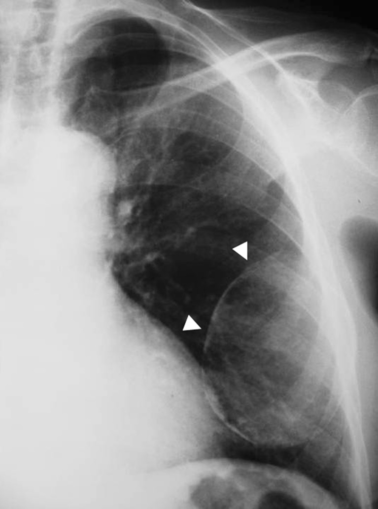
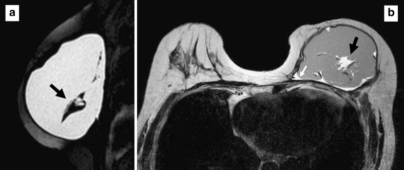
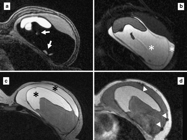
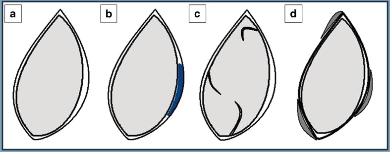
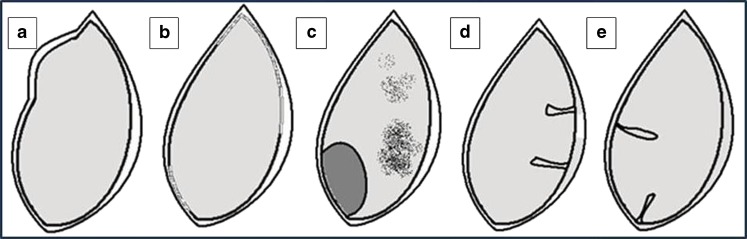
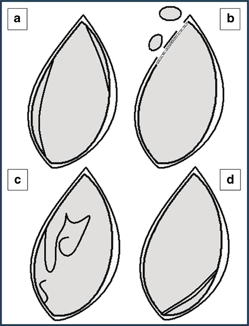
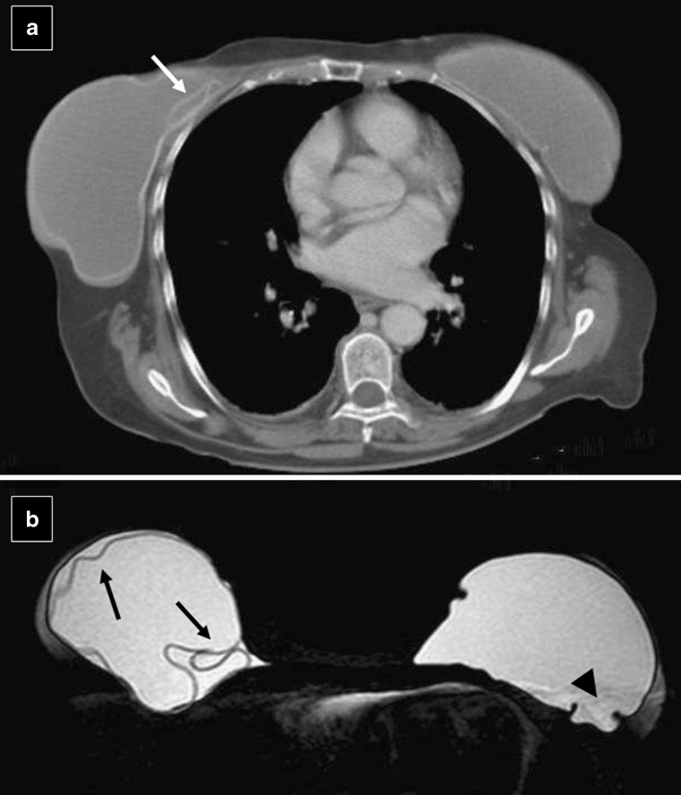
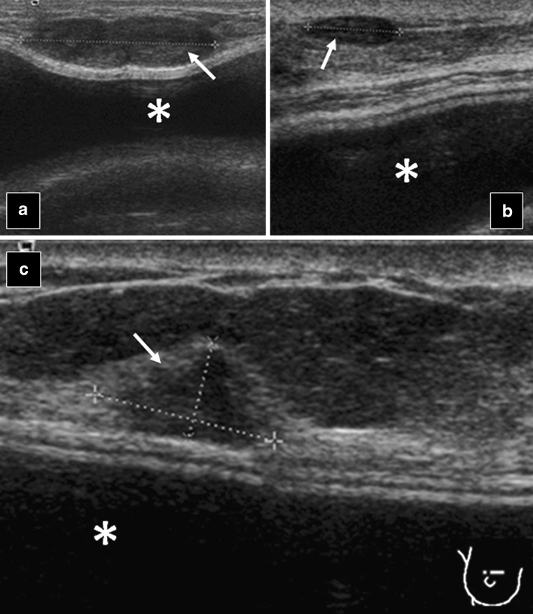
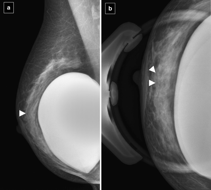
The number of women with breast implants is increasing. Radiologists must be familiar with the normal and abnormal findings of common implants. Implant rupture is a well-known complication after surgery and is the main cause of implant removal. Although mammography and ultrasonography are the standard first steps in the diagnostic workup, magnetic resonance imaging (MRI) is the most useful imaging modality for the characterisation of breast implants because of its high spatial resolution and contrast between implants and soft tissues and absence of ionising radiation. MRI has the highest sensitivity and specificity for implant rupture, thanks to its sequences that can suppress or emphasise the signal from silicone. Regardless of the technique used, the overall aim of imaging breast implants is to provide essential information about tissue and prosthesis integrity, detect implant abnormalities and detect breast diseases unrelated to implants, such as breast cancer.
接受隆胸手术的女性数量在不断增加。放射科医生必须熟悉常见植入物的正常和异常表现。植入物破裂是手术后一种众所周知的并发症,也是移除植入物的主要原因。尽管乳房X线摄影和超声检查是诊断检查的标准第一步,但磁共振成像(MRI)因其高空间分辨率、植入物与软组织之间的对比度以及无电离辐射,是用于表征乳房植入物最有用的成像方式。由于其能够抑制或增强硅胶信号的序列,MRI对植入物破裂具有最高的敏感性和特异性。无论使用何种技术,对乳房植入物进行成像的总体目标是提供有关组织和假体完整性的基本信息,检测植入物异常,并检测与植入物无关的乳腺疾病,如乳腺癌。