Laboratory of Malaria Immunobiology, Singapore Immunology Network (SIgN), Agency for Science, Technology and Research(A*STAR), Biopolis, Singapore.
Sci Rep. 2011;1:118. doi: 10.1038/srep00118. Epub 2011 Oct 14.
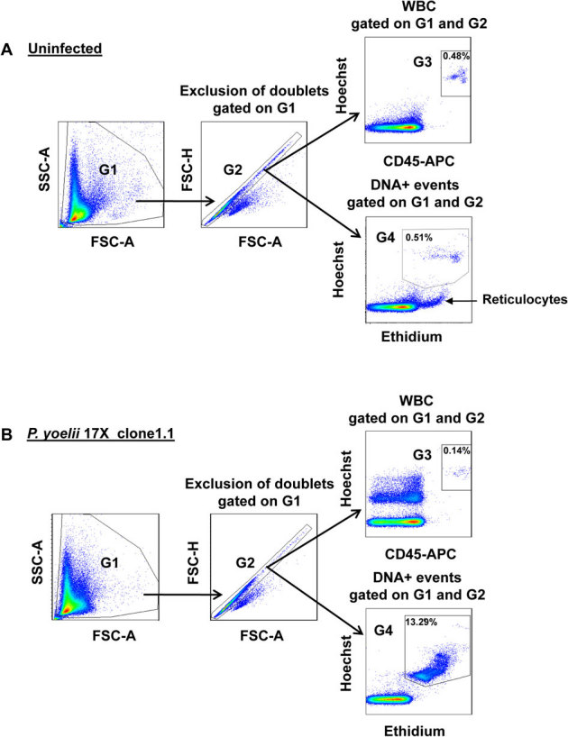
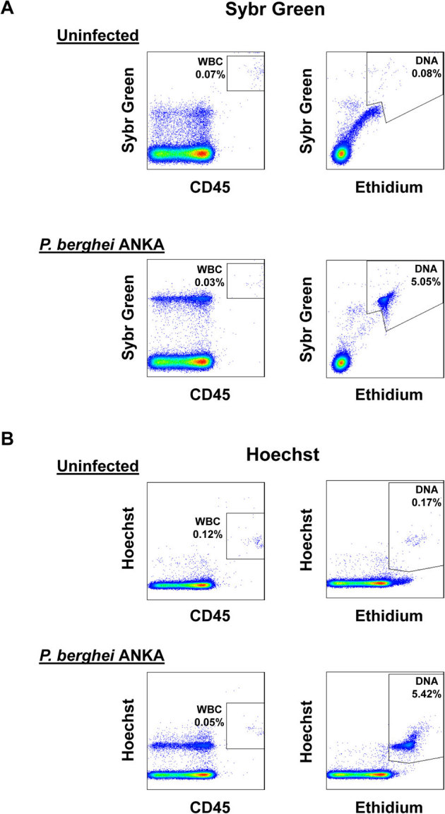
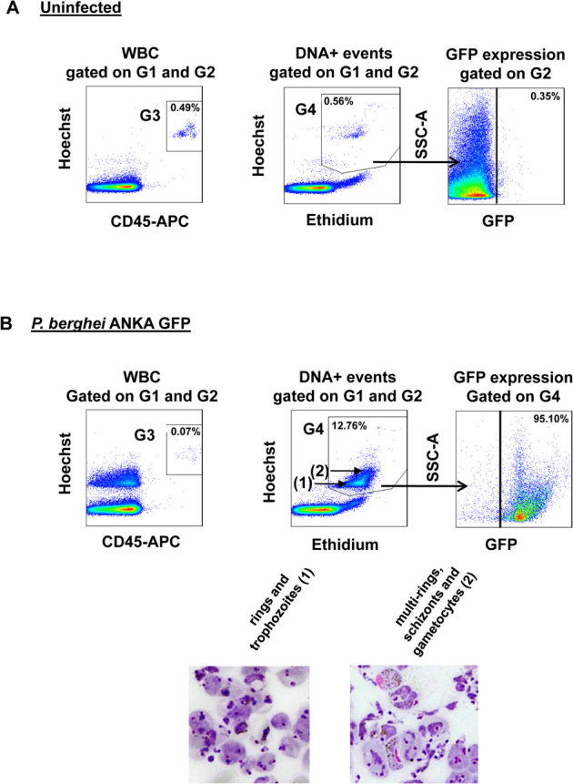
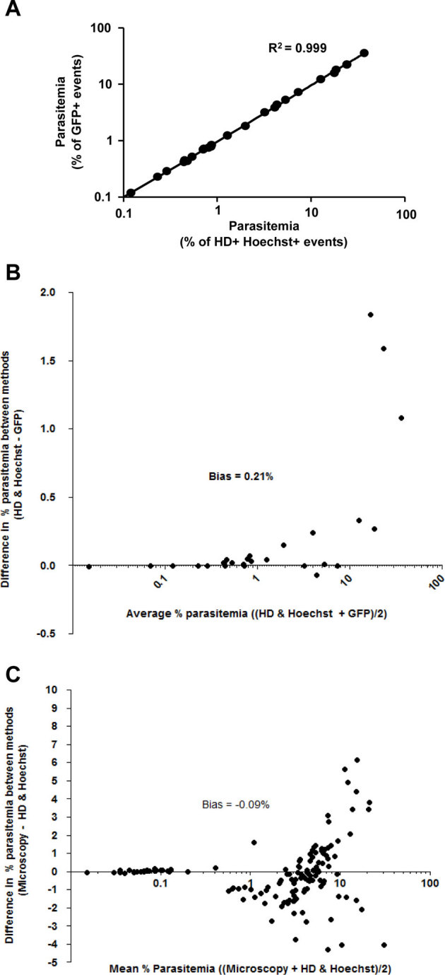
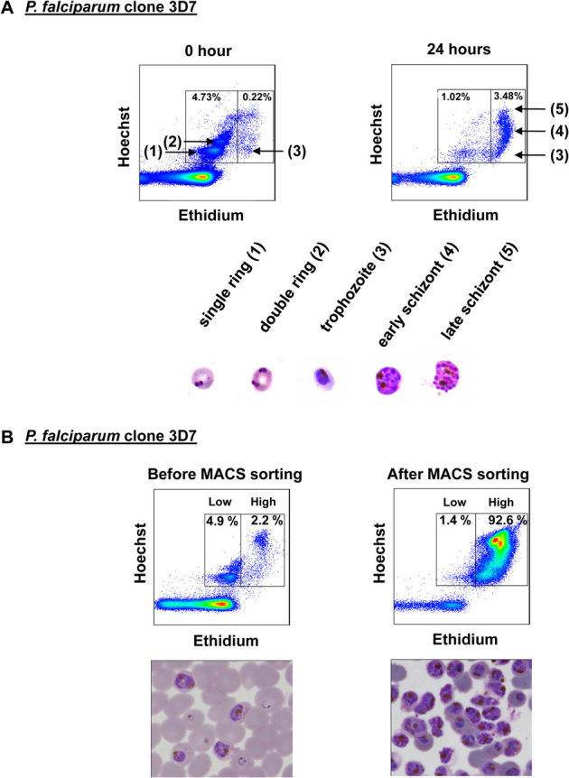
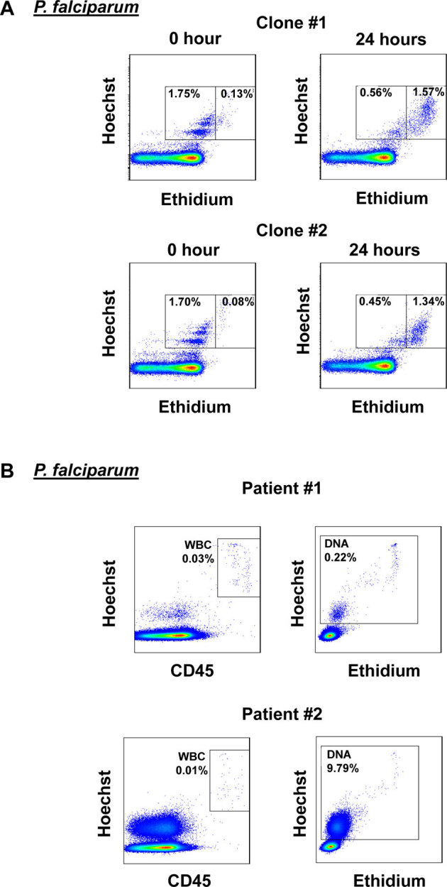
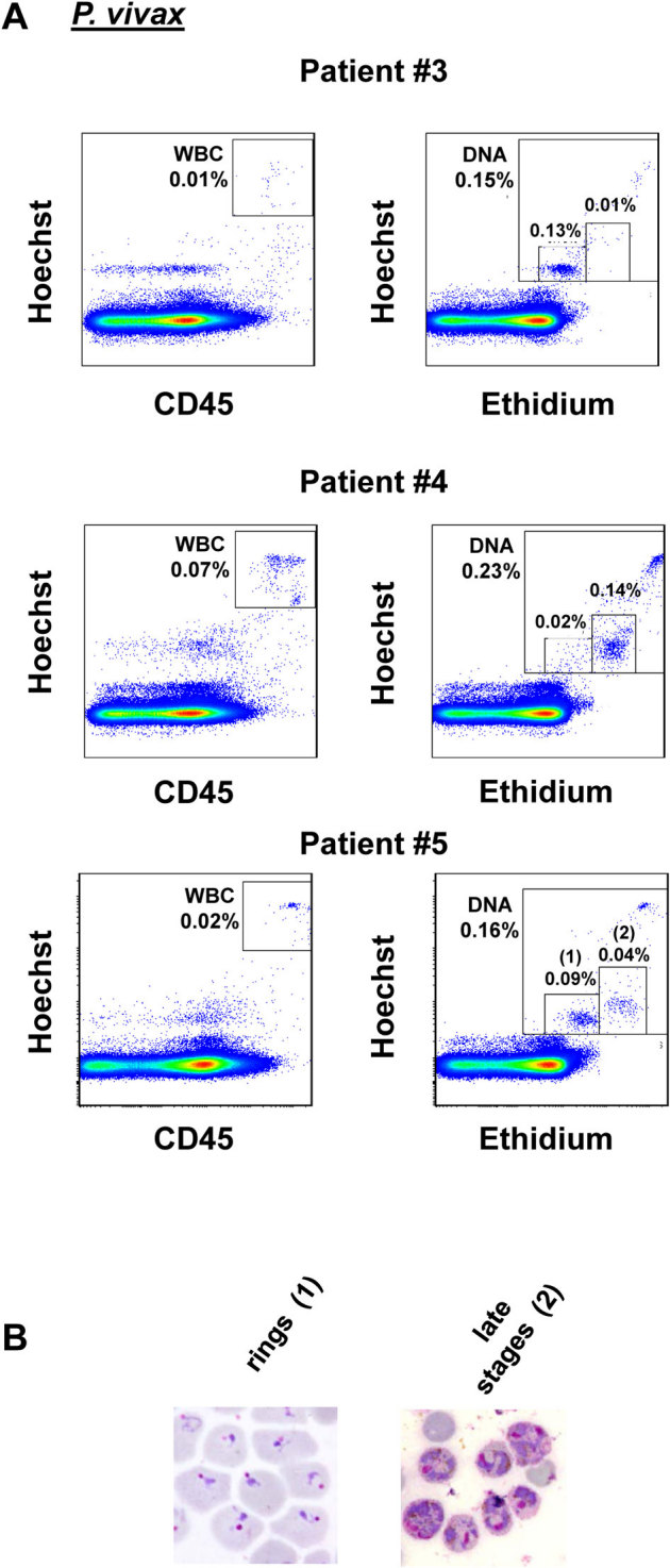
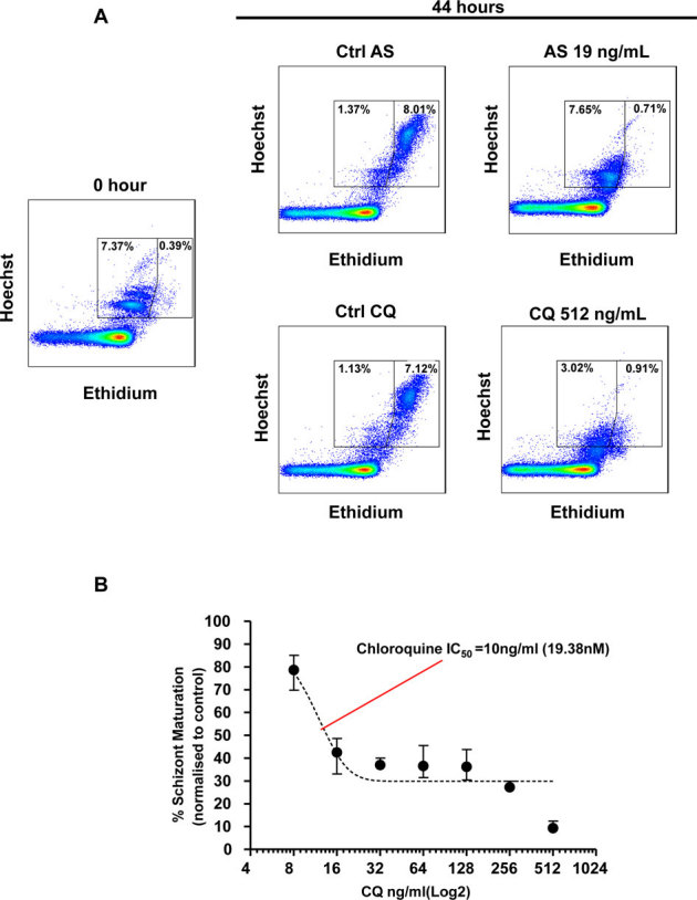
Microscopic examination of Giemsa-stained thin blood smears remains the gold standard method used to quantify and stage malaria parasites. However, this technique is tedious, and requires trained microscopists. We have developed a fast and simple flow cytometry method to quantify and stage, various malaria parasites in red blood cells in whole blood or in vitro cultured Plasmodium falciparum. The parasites were stained with dihydroethidium and Hoechst 33342 or SYBR Green I and leukocytes were identified with an antibody against CD45. Depending on the DNA stains used, samples were analyzed using different models of flow cytometers. This protocol, which does not require any washing steps, allows infected red blood cells to be distinguished from leukocytes, as well as allowing non-infected reticulocytes and normocytes to be identified. It also allows assessing the proportion of parasites at different developmental stages. Lastly, we demonstrate how this technique can be applied to antimalarial drug testing.
吉姆萨染色薄血涂片的显微镜检查仍然是定量和分期疟原虫的金标准方法。然而,这种技术繁琐,需要经过培训的显微镜专家。我们开发了一种快速而简单的流式细胞术方法,用于定量和分期各种疟原虫在全血或体外培养的恶性疟原虫中的红细胞。用二氢乙啶和 Hoechst 33342 或 SYBR Green I 对寄生虫进行染色,并用针对 CD45 的抗体识别白细胞。根据使用的 DNA 染色剂,使用不同型号的流式细胞仪分析样本。该方案不需要任何洗涤步骤,允许区分感染的红细胞与白细胞,同时还允许识别未感染的网织红细胞和正常红细胞。它还可以评估不同发育阶段的寄生虫比例。最后,我们展示了如何将该技术应用于抗疟药物测试。