• 文献检索
  • 文档翻译
  • 深度研究
  • 学术资讯
  • Suppr Zotero 插件Zotero 插件
  • 邀请有礼
  • 套餐&价格
  • 历史记录
应用&插件
Suppr Zotero 插件Zotero 插件浏览器插件Mac 客户端Windows 客户端微信小程序
定价
高级版会员购买积分包购买API积分包
服务
文献检索文档翻译深度研究API 文档MCP 服务
关于我们
关于 Suppr公司介绍联系我们用户协议隐私条款
关注我们

Suppr 超能文献

核心技术专利:CN118964589B侵权必究
粤ICP备2023148730 号-1Suppr @ 2026

文献检索

告别复杂PubMed语法,用中文像聊天一样搜索,搜遍4000万医学文献。AI智能推荐,让科研检索更轻松。

立即免费搜索

文件翻译

保留排版,准确专业,支持PDF/Word/PPT等文件格式,支持 12+语言互译。

免费翻译文档

深度研究

AI帮你快速写综述,25分钟生成高质量综述,智能提取关键信息,辅助科研写作。

立即免费体验

房间隔缺损的三维经食管超声心动图评估:图文综述。

Three-dimensional trans-esophageal Echocardiographic Evaluation of Atrial Septal Defects: A Pictorial Essay.

作者信息

Sharma Vinay K, Radhakrishnan S, Shrivastava S

机构信息

Fortis Escorts Heart Institute and Research Centre Ltd., Okhla road, New Delhi-110025, India.

出版信息

Images Paediatr Cardiol. 2011 Jul;13(3):1-18.

PMID:22368571
原文链接:https://pmc.ncbi.nlm.nih.gov/articles/PMC3232598/
Abstract

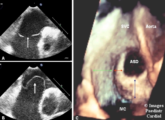
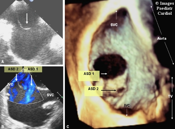
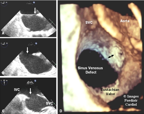
This pictorial assay illustrates the methodology of evaluating the atrial septal defects by three dimensional transesophageal echocardiography with the help of representative images. The article starts by discussing the technical details of how to acquire and crop the dataset to reconstruct the transesophageal three dimensional echocardiographic images of the inter atrial septum. Next, the anatomical details of the normal inter atrial septum are illustrated, followed by representative examples of all the possible defects of inter atrial septum. All the images have been reproduced in a uniform pattern which is similar to the view of the inter atrial septum that is seen in the real life situation by the surgeon.

摘要

本图像分析借助代表性图像阐述了三维经食管超声心动图评估房间隔缺损的方法。文章首先讨论了获取和裁剪数据集以重建房间隔经食管三维超声心动图图像的技术细节。接下来,展示了正常房间隔的解剖细节,随后是房间隔所有可能缺损的代表性实例。所有图像均以统一模式呈现,类似于外科医生在实际情况中看到的房间隔视图。

https://cdn.ncbi.nlm.nih.gov/pmc/blobs/8bbc/3232598/12313e5ca18b/IPC-13-1-g014.jpg
https://cdn.ncbi.nlm.nih.gov/pmc/blobs/8bbc/3232598/212fef3b5e43/IPC-13-1-g001.jpg
https://cdn.ncbi.nlm.nih.gov/pmc/blobs/8bbc/3232598/38d9791f1b3d/IPC-13-1-g002.jpg
https://cdn.ncbi.nlm.nih.gov/pmc/blobs/8bbc/3232598/82dfd8e81e3a/IPC-13-1-g003.jpg
https://cdn.ncbi.nlm.nih.gov/pmc/blobs/8bbc/3232598/9526338867a1/IPC-13-1-g004.jpg
https://cdn.ncbi.nlm.nih.gov/pmc/blobs/8bbc/3232598/775a97e66d2d/IPC-13-1-g005.jpg
https://cdn.ncbi.nlm.nih.gov/pmc/blobs/8bbc/3232598/f347030a1aaa/IPC-13-1-g006.jpg
https://cdn.ncbi.nlm.nih.gov/pmc/blobs/8bbc/3232598/c0b2c4d995f0/IPC-13-1-g007.jpg
https://cdn.ncbi.nlm.nih.gov/pmc/blobs/8bbc/3232598/8b9c919a730c/IPC-13-1-g008.jpg
https://cdn.ncbi.nlm.nih.gov/pmc/blobs/8bbc/3232598/90480dc418f2/IPC-13-1-g009.jpg
https://cdn.ncbi.nlm.nih.gov/pmc/blobs/8bbc/3232598/c1c57e4ba1a3/IPC-13-1-g010.jpg
https://cdn.ncbi.nlm.nih.gov/pmc/blobs/8bbc/3232598/0199906b66ea/IPC-13-1-g011.jpg
https://cdn.ncbi.nlm.nih.gov/pmc/blobs/8bbc/3232598/9fcb8cfa708c/IPC-13-1-g012.jpg
https://cdn.ncbi.nlm.nih.gov/pmc/blobs/8bbc/3232598/bfdefa0ef5e1/IPC-13-1-g013.jpg
https://cdn.ncbi.nlm.nih.gov/pmc/blobs/8bbc/3232598/12313e5ca18b/IPC-13-1-g014.jpg

相似文献

1
Three-dimensional trans-esophageal Echocardiographic Evaluation of Atrial Septal Defects: A Pictorial Essay.房间隔缺损的三维经食管超声心动图评估:图文综述。
Images Paediatr Cardiol. 2011 Jul;13(3):1-18.
2
Three-dimensional ultrasound imaging of the atrial septum: normal and pathologic anatomy.
J Am Coll Cardiol. 1993 Nov 15;22(6):1673-8. doi: 10.1016/0735-1097(93)90594-q.
3
Transesophageal echocardiographic evaluation of atrial septal defects--how to explore the third dimension?经食管超声心动图对房间隔缺损的评估——如何探索三维空间?
Indian Heart J. 2009 Jul-Aug;61(4):328-34.
4
Three-dimensional transesophageal echocardiography of the atrial septal defects.房间隔缺损的三维经食管超声心动图检查
Cardiovasc Ultrasound. 2008 Jul 18;6:38. doi: 10.1186/1476-7120-6-38.
5
Single and two dimensional echocardiographic features of the interatrial septum in normal subjects and patients with an atrial septal defect.正常受试者和房间隔缺损患者房间隔的一维及二维超声心动图特征
Am J Cardiol. 1979 Apr;43(4):816-9. doi: 10.1016/0002-9149(79)90083-3.
6
Imaging the atrial septum using real-time three-dimensional transesophageal echocardiography: technical tips, normal anatomy, and its role in transseptal puncture.实时三维经食管超声心动图成像技术在房间隔中的应用:技术要点、正常解剖结构及其在房间隔穿刺中的作用。
J Am Soc Echocardiogr. 2011 Jun;24(6):593-9. doi: 10.1016/j.echo.2011.01.022. Epub 2011 Mar 9.
7
[Surgical evaluation of transthoracic tridimensional echocardiography in the anatomic study of atrial septal defect].经胸三维超声心动图在房间隔缺损解剖学研究中的外科评估
Arch Mal Coeur Vaiss. 1999 May;92(5):573-80.
8
Imaging atrial septal defects by real-time three-dimensional transesophageal echocardiography: step-by-step approach.实时三维经食管超声心动图成像房间隔缺损:一步一步的方法。
J Am Soc Echocardiogr. 2010 Nov;23(11):1128-35. doi: 10.1016/j.echo.2010.08.008. Epub 2010 Sep 15.
9
Three-dimensional echocardiography of the atrial septum.房间隔的三维超声心动图
Echocardiography. 2001 Jul;18(5):433-43. doi: 10.1046/j.1540-8175.2001.00433.x.
10
Delineation of site, relative size and dynamic geometry of atrial septal defects by real-time three-dimensional echocardiography.实时三维超声心动图对房间隔缺损部位、相对大小及动态几何学特征的描绘
J Am Coll Cardiol. 1995 Feb;25(2):482-90. doi: 10.1016/0735-1097(94)00372-w.

引用本文的文献

1
Assessment of Value of Three Dimensional Transesophageal Echocardiography versus Conventional Two Dimensional Transesophageal Echocardiography in Guiding Transcatheter Closure of Atrial Septal Defects and Patent Foramen Ovale.三维经食管超声心动图与传统二维经食管超声心动图在指导房间隔缺损和卵圆孔未闭经导管封堵术中的价值评估
J Saudi Heart Assoc. 2022 Jul 6;34(1):85-99. doi: 10.37616/2212-5043.1303. eCollection 2022.

本文引用的文献

1
Incidence and predictors of atrial septal defect after percutaneous transvenous mitral commissurotomy - a transesophageal echocardiographic study of 209 cases.经皮经静脉二尖瓣交界切开术后房间隔缺损的发生率及预测因素——一项对209例患者的经食管超声心动图研究
Echocardiography. 2013 Feb;30(2):127-30. doi: 10.1111/echo.12025. Epub 2012 Dec 12.
2
Transesophageal echocardiographic evaluation of atrial septal defects--how to explore the third dimension?经食管超声心动图对房间隔缺损的评估——如何探索三维空间?
Indian Heart J. 2009 Jul-Aug;61(4):328-34.
3
Interventional cardiology and cardiac surgery in India.
Heart. 2008 Mar;94(3):268-74. doi: 10.1136/hrt.2007.133041.
4
Echocardiographic anatomy of atrial septal defect: "nomenclature of the rims".房间隔缺损的超声心动图解剖:“边缘的命名”
Indian Heart J. 2003 Jan-Feb;55(1):88-9.
5
Morphological variations of secundum-type atrial septal defects: feasibility for percutaneous closure using Amplatzer septal occluders.继发孔型房间隔缺损的形态学变异:使用Amplatzer房间隔封堵器进行经皮封堵的可行性
Catheter Cardiovasc Interv. 2001 Jul;53(3):386-91. doi: 10.1002/ccd.1187.
6
Dynamic morphology of the secundum atrial septal defect evaluated by three dimensional transoesophageal echocardiography.经三维经食管超声心动图评估的继发孔型房间隔缺损的动态形态学
Heart. 2000 Jun;83(6):673-7. doi: 10.1136/heart.83.6.673.
7
Transcatheter closure of multiple atrial septal defects. Initial results and value of two- and three-dimensional transoesophageal echocardiography.经导管封堵多发性房间隔缺损。二维和三维经食管超声心动图的初步结果及价值。
Eur Heart J. 2000 Jun;21(11):941-7. doi: 10.1053/euhj.1999.1909.
8
A new technique for sizing of atrial septal defects.一种用于测量房间隔缺损大小的新技术。
Catheter Cardiovasc Interv. 1999 Jan;46(1):51-7. doi: 10.1002/(SICI)1522-726X(199901)46:1<51::AID-CCD14>3.0.CO;2-M.
9
Assessment of atrial septal defect morphology by transthoracic three dimensional echocardiography using standard grey scale and Doppler myocardial imaging techniques: comparison with magnetic resonance imaging and intraoperative findings.使用标准灰阶和多普勒心肌成像技术经胸三维超声心动图评估房间隔缺损形态:与磁共振成像及术中发现的比较
Heart. 1997 Oct;78(4):382-9. doi: 10.1136/hrt.78.4.382.
10
Anatomical criteria for the diagnosis of sinus venosus defects.诊断静脉窦缺损的解剖学标准。
Heart. 1997 Sep;78(3):298-304. doi: 10.1136/hrt.78.3.298.