• 文献检索
  • 文档翻译
  • 深度研究
  • 学术资讯
  • Suppr Zotero 插件Zotero 插件
  • 邀请有礼
  • 套餐&价格
  • 历史记录
应用&插件
Suppr Zotero 插件Zotero 插件浏览器插件Mac 客户端Windows 客户端微信小程序
定价
高级版会员购买积分包购买API积分包
服务
文献检索文档翻译深度研究API 文档MCP 服务
关于我们
关于 Suppr公司介绍联系我们用户协议隐私条款
关注我们

Suppr 超能文献

核心技术专利:CN118964589B侵权必究
粤ICP备2023148730 号-1Suppr @ 2026

文献检索

告别复杂PubMed语法,用中文像聊天一样搜索,搜遍4000万医学文献。AI智能推荐,让科研检索更轻松。

立即免费搜索

文件翻译

保留排版,准确专业,支持PDF/Word/PPT等文件格式,支持 12+语言互译。

免费翻译文档

深度研究

AI帮你快速写综述,25分钟生成高质量综述,智能提取关键信息,辅助科研写作。

立即免费体验

抑制转化生长因子激活激酶 1(TAK1)可阻断并逆转间皮细胞的上皮间质转化。

Inhibition of transforming growth factor-activated kinase 1 (TAK1) blocks and reverses epithelial to mesenchymal transition of mesothelial cells.

机构信息

Integrin Signaling Laboratory, Department of Vascular Biology and Inflammation, Centro Nacional de Investigaciones Cardiovasculares, Madrid, Spain.

出版信息

PLoS One. 2012;7(2):e31492. doi: 10.1371/journal.pone.0031492. Epub 2012 Feb 27.

DOI:10.1371/journal.pone.0031492
PMID:22384029
原文链接:https://pmc.ncbi.nlm.nih.gov/articles/PMC3288041/
Abstract

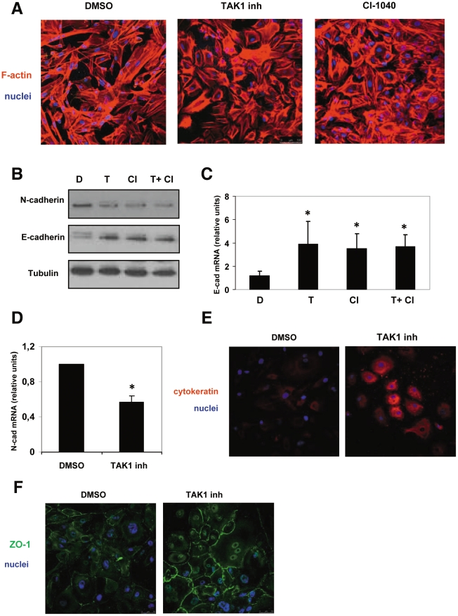
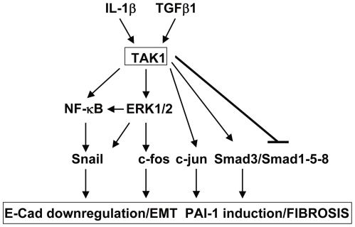
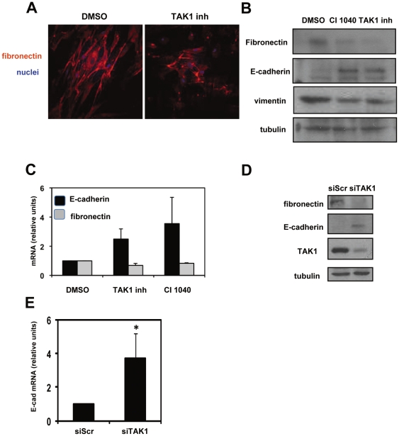
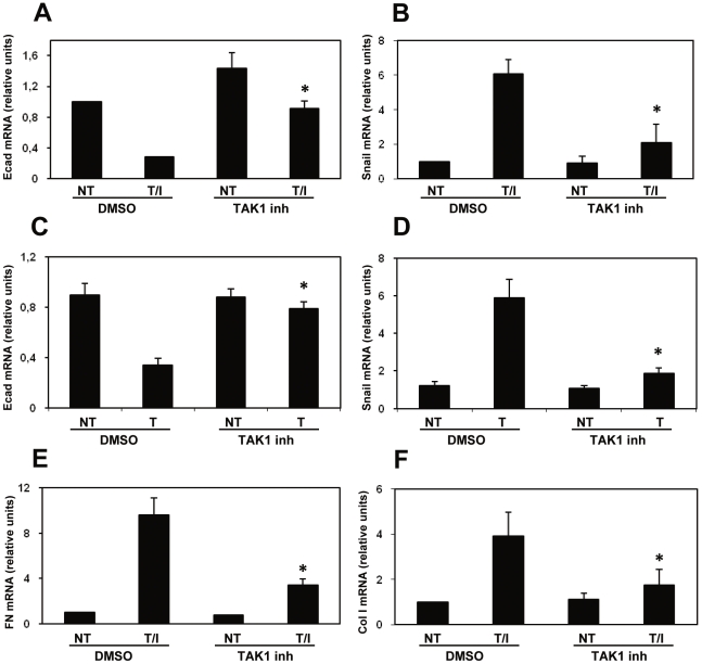
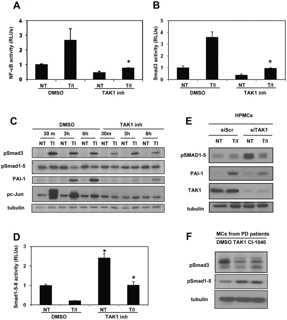
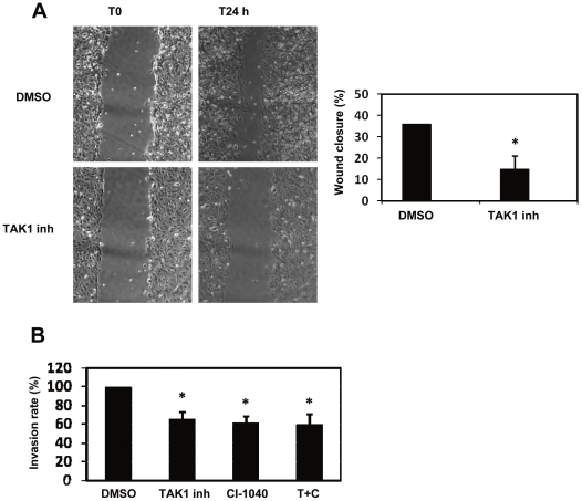
Peritoneal fibrosis is a frequent complication of peritoneal dialysis following repeated low grade inflammatory and pro-fibrotic insults. This pathological process may lead to ultrafiltration failure and eventually to the discontinuing of the therapy. Fibrosis is linked to epithelial to mesenchymal transition (EMT) of the peritoneal mesothelial cells, which acquire invasive and fibrogenic abilities. Here, we analyzed the role of the transforming growth factor-activated kinase-1 (TAK1) in the EMT of primary mesothelial cells from human peritoneum. The inhibition of TAK1 in mesenchymal-like mesothelial cells from the effluents of patients undergoing peritoneal dialysis led to the reacquisition of the apical to basolateral polarity, to increased expression of epithelial and to down-regulation of mesenchymal markers. TAK1 inhibition also resulted in decreased migratory/invasive abilities of effluent-derived mesothelial cells. Simultaneous inhibition of ERK1/2 and TAK1 pathways did not lead to an additive effect in the reacquisition of the epithelial phenotype. Inhibition of TAK1 also blocked EMT in vitro and reduced the levels of PAI-1, which is involved in fibrosis and invasion. Analysis of signalling pathways downstream of TAK1 involved in EMT induction, showed that TAK1 inhibition reduced the transcriptional activity of NF-κB and Smad3, as well as the phosphorylation of c-jun, while enhancing Smad1-5-8 activity. These results demonstrate that TAK1 is a cross-point in a network including different pro-EMT transcription factors, such as NF-κB, Snail, AP-1 and Smads. The identification of TAK1 as a main biochemical mediator of EMT and fibrosis in mesothelial cells from human peritoneum and the study of signalling pathways induced by its activity may be relevant in the design of new therapies aimed to counteract peritoneal fibrosis.

摘要

腹膜纤维化是腹膜透析后反复发生低度炎症和促纤维化损伤的常见并发症。这一病理过程可导致超滤衰竭,并最终导致治疗中断。纤维化与腹膜间皮细胞的上皮间质转化(EMT)有关,间皮细胞获得侵袭和纤维化能力。在这里,我们分析了转化生长因子激活激酶 1(TAK1)在人腹膜间皮细胞 EMT 中的作用。在接受腹膜透析的患者流出物中的间充质样间皮细胞中抑制 TAK1 可导致顶端到基底外侧极性的重新获得、上皮标志物的表达增加和间充质标志物的下调。TAK1 抑制也导致流出物衍生的间皮细胞迁移/侵袭能力降低。ERK1/2 和 TAK1 通路的同时抑制在获得上皮表型方面没有叠加效应。TAK1 抑制还阻断了 EMT,并降低了参与纤维化和侵袭的 PAI-1 水平。对 TAK1 下游参与 EMT 诱导的信号通路的分析表明,TAK1 抑制减少了 NF-κB 和 Smad3 的转录活性以及 c-jun 的磷酸化,同时增强了 Smad1-5-8 的活性。这些结果表明 TAK1 是一个包含不同 EMT 转录因子(如 NF-κB、Snail、AP-1 和 Smads)的网络中的交汇点。确定 TAK1 是人类腹膜间皮细胞 EMT 和纤维化的主要生化介质,以及研究其活性诱导的信号通路,可能与设计旨在对抗腹膜纤维化的新疗法有关。

https://cdn.ncbi.nlm.nih.gov/pmc/blobs/ddd1/3288041/b820a73ccc67/pone.0031492.g006.jpg
https://cdn.ncbi.nlm.nih.gov/pmc/blobs/ddd1/3288041/9f9005eab810/pone.0031492.g001.jpg
https://cdn.ncbi.nlm.nih.gov/pmc/blobs/ddd1/3288041/cd1b37f2922d/pone.0031492.g002.jpg
https://cdn.ncbi.nlm.nih.gov/pmc/blobs/ddd1/3288041/c8d85bffc661/pone.0031492.g003.jpg
https://cdn.ncbi.nlm.nih.gov/pmc/blobs/ddd1/3288041/5bb7089d9e65/pone.0031492.g004.jpg
https://cdn.ncbi.nlm.nih.gov/pmc/blobs/ddd1/3288041/0c7b77017f1e/pone.0031492.g005.jpg
https://cdn.ncbi.nlm.nih.gov/pmc/blobs/ddd1/3288041/b820a73ccc67/pone.0031492.g006.jpg

相似文献

1
Inhibition of transforming growth factor-activated kinase 1 (TAK1) blocks and reverses epithelial to mesenchymal transition of mesothelial cells.抑制转化生长因子激活激酶 1(TAK1)可阻断并逆转间皮细胞的上皮间质转化。
PLoS One. 2012;7(2):e31492. doi: 10.1371/journal.pone.0031492. Epub 2012 Feb 27.
2
Curcumin suppresses epithelial-to-mesenchymal transition of peritoneal mesothelial cells (HMrSV5) through regulation of transforming growth factor-activated kinase 1 (TAK1).姜黄素通过调节转化生长因子激活激酶 1(TAK1)抑制腹膜间皮细胞(HMrSV5)的上皮间质转化。
Cell Mol Biol Lett. 2019 May 22;24:32. doi: 10.1186/s11658-019-0157-x. eCollection 2019.
3
p38 maintains E-cadherin expression by modulating TAK1-NF-kappa B during epithelial-to-mesenchymal transition.p38 通过调节上皮-间充质转化过程中的 TAK1-NF-κB 来维持 E-钙黏蛋白的表达。
J Cell Sci. 2010 Dec 15;123(Pt 24):4321-31. doi: 10.1242/jcs.071647. Epub 2010 Nov 23.
4
Histone acetyltransferase inhibitor C646 reverses epithelial to mesenchymal transition of human peritoneal mesothelial cells via blocking TGF-β1/Smad3 signaling pathway in vitro.组蛋白乙酰转移酶抑制剂C646在体外通过阻断TGF-β1/Smad3信号通路逆转人腹膜间皮细胞的上皮-间质转化。
Int J Clin Exp Pathol. 2015 Mar 1;8(3):2746-54. eCollection 2015.
5
Arctigenin alleviates TGF-β1-induced epithelial-mesenchymal transition and PAI-1 expression via AMPK/NF-κB pathway in peritoneal mesothelial cells.原花青素通过 AMPK/NF-κB 通路减轻 TGF-β1 诱导的腹膜间皮细胞上皮-间充质转化和 PAI-1 表达。
Biochem Biophys Res Commun. 2019 Dec 3;520(2):413-419. doi: 10.1016/j.bbrc.2019.09.130. Epub 2019 Oct 11.
6
Curcumin ameliorates peritoneal fibrosis via inhibition of transforming growth factor-activated kinase 1 (TAK1) pathway in a rat model of peritoneal dialysis.姜黄素通过抑制转化生长因子激活激酶 1(TAK1)通路减轻腹膜透析大鼠模型的腹膜纤维化。
BMC Complement Altern Med. 2019 Oct 23;19(1):280. doi: 10.1186/s12906-019-2702-6.
7
Epithelial-to-mesenchymal transition of peritoneal mesothelial cells is regulated by an ERK/NF-kappaB/Snail1 pathway.腹膜间皮细胞的上皮-间质转化由ERK/NF-κB/Snail1信号通路调控。
Dis Model Mech. 2008 Nov-Dec;1(4-5):264-74. doi: 10.1242/dmm.001321. Epub 2008 Oct 28.
8
Nitro-oleic acid inhibits the high glucose-induced epithelial-mesenchymal transition in peritoneal mesothelial cells and attenuates peritoneal fibrosis.硝异山梨醇抑制高糖诱导的腹膜间皮细胞上皮间质转化并减轻腹膜纤维化。
Am J Physiol Renal Physiol. 2020 Feb 1;318(2):F457-F467. doi: 10.1152/ajprenal.00425.2019. Epub 2019 Nov 25.
9
Autophagy promotes fibrosis and apoptosis in the peritoneum during long-term peritoneal dialysis.自噬促进长期腹膜透析中腹膜纤维化和细胞凋亡。
J Cell Mol Med. 2018 Feb;22(2):1190-1201. doi: 10.1111/jcmm.13393. Epub 2017 Oct 27.
10
The critical role of TAK1 in accentuated epithelial to mesenchymal transition in obliterative bronchiolitis after lung transplantation.TAK1 在肺移植后闭塞性细支气管炎中上皮间质转化增强中的关键作用。
Am J Pathol. 2012 Jun;180(6):2293-308. doi: 10.1016/j.ajpath.2012.02.022. Epub 2012 Apr 21.

引用本文的文献

1
Detection of Carcinoma-Associated Fibroblasts Derived from Mesothelial Cells via Mesothelial-to-Mesenchymal Transition in Primary Ovarian Carcinomas.通过原发性卵巢癌中上皮-间质转化检测源自间皮细胞的癌相关成纤维细胞
Cancers (Basel). 2024 Jul 29;16(15):2697. doi: 10.3390/cancers16152697.
2
HDAC1/2 control mesothelium/ovarian cancer adhesive interactions impacting on Talin-1-α5β1-integrin-mediated actin cytoskeleton and extracellular matrix protein remodeling.组蛋白去乙酰化酶 1/2 控制间皮/卵巢癌细胞黏附相互作用,影响 Talin-1-α5β1-整合素介导的肌动蛋白细胞骨架和细胞外基质蛋白重塑。
J Exp Clin Cancer Res. 2024 Jan 23;43(1):27. doi: 10.1186/s13046-023-02930-8.
3

本文引用的文献

1
Influence of bicarbonate/low-GDP peritoneal dialysis fluid (BicaVera) on in vitro and ex vivo epithelial-to-mesenchymal transition of mesothelial cells.碳酸氢盐/低 GDP 腹膜透析液(BicaVera)对间皮细胞体外和体内上皮间质转化的影响。
Perit Dial Int. 2012 May-Jun;32(3):292-304. doi: 10.3747/pdi.2010.00315. Epub 2012 Jan 3.
2
p38 maintains E-cadherin expression by modulating TAK1-NF-kappa B during epithelial-to-mesenchymal transition.p38 通过调节上皮-间充质转化过程中的 TAK1-NF-κB 来维持 E-钙黏蛋白的表达。
J Cell Sci. 2010 Dec 15;123(Pt 24):4321-31. doi: 10.1242/jcs.071647. Epub 2010 Nov 23.
3
Glucocorticoid induces mesenchymal-to-epithelial transition and inhibits TGF-β1-induced epithelial-to-mesenchymal transition and cell migration.
Chronic treatment with TNF-α, alone and in combination with Takinib, SB203580 and metformin induce cell death in breast cancer.
单独使用TNF-α以及与塔替尼、SB203580和二甲双胍联合进行长期治疗可诱导乳腺癌细胞死亡。
Heliyon. 2023 Oct 16;9(11):e21060. doi: 10.1016/j.heliyon.2023.e21060. eCollection 2023 Nov.
4
PCB: A pseudotemporal causality-based Bayesian approach to identify EMT-associated regulatory relationships of AS events and RBPs during breast cancer progression.PCB:一种基于伪时间因果关系的贝叶斯方法,用于识别乳腺癌进展过程中 AS 事件和 RBPs 的 EMT 相关调控关系。
PLoS Comput Biol. 2023 Mar 17;19(3):e1010939. doi: 10.1371/journal.pcbi.1010939. eCollection 2023 Mar.
5
Regulation of Mesothelial Cell Fate during Development and Human Diseases.胚胎发育和人类疾病过程中间皮细胞命运的调控。
Int J Mol Sci. 2022 Oct 8;23(19):11960. doi: 10.3390/ijms231911960.
6
Post-Surgical Peritoneal Scarring and Key Molecular Mechanisms.术后腹膜瘢痕形成及关键分子机制。
Biomolecules. 2021 May 5;11(5):692. doi: 10.3390/biom11050692.
7
Mechanisms of Peritoneal Fibrosis: Focus on Immune Cells-Peritoneal Stroma Interactions.腹膜纤维化的机制:聚焦免疫细胞-腹膜基质细胞相互作用。
Front Immunol. 2021 Mar 29;12:607204. doi: 10.3389/fimmu.2021.607204. eCollection 2021.
8
The Epithelial-to-Mesenchymal Transition as a Possible Therapeutic Target in Fibrotic Disorders.上皮-间质转化作为纤维化疾病潜在的治疗靶点
Front Cell Dev Biol. 2020 Dec 21;8:607483. doi: 10.3389/fcell.2020.607483. eCollection 2020.
9
IL-17A as a Potential Therapeutic Target for Patients on Peritoneal Dialysis.白细胞介素-17A 作为腹膜透析患者的潜在治疗靶点。
Biomolecules. 2020 Sep 24;10(10):1361. doi: 10.3390/biom10101361.
10
Curcumin ameliorates peritoneal fibrosis via inhibition of transforming growth factor-activated kinase 1 (TAK1) pathway in a rat model of peritoneal dialysis.姜黄素通过抑制转化生长因子激活激酶 1(TAK1)通路减轻腹膜透析大鼠模型的腹膜纤维化。
BMC Complement Altern Med. 2019 Oct 23;19(1):280. doi: 10.1186/s12906-019-2702-6.
糖皮质激素诱导间充质-上皮转化,抑制 TGF-β1 诱导的上皮-间充质转化和细胞迁移。
FEBS Lett. 2010 Nov 19;584(22):4646-54. doi: 10.1016/j.febslet.2010.10.038. Epub 2010 Oct 26.
4
c-Jun N-terminal kinase 1 promotes transforming growth factor-β1-induced epithelial-to-mesenchymal transition via control of linker phosphorylation and transcriptional activity of Smad3.c-Jun N-末端激酶 1 通过控制链接子磷酸化和 Smad3 的转录活性促进转化生长因子-β1 诱导的上皮-间充质转化。
Am J Respir Cell Mol Biol. 2011 Apr;44(4):571-81. doi: 10.1165/rcmb.2009-0282OC. Epub 2010 Jun 25.
5
The pathophysiology of the peritoneal membrane.腹膜的病理生理学。
J Am Soc Nephrol. 2010 Jul;21(7):1077-85. doi: 10.1681/ASN.2009070694. Epub 2010 May 6.
6
Disruption of TAK1 in hepatocytes causes hepatic injury, inflammation, fibrosis, and carcinogenesis.TAK1 在肝细胞中的缺失会导致肝损伤、炎症、纤维化和癌变。
Proc Natl Acad Sci U S A. 2010 Jan 12;107(2):844-9. doi: 10.1073/pnas.0909781107. Epub 2009 Dec 18.
7
BMP-7 blocks mesenchymal conversion of mesothelial cells and prevents peritoneal damage induced by dialysis fluid exposure.BMP-7 可阻止间皮细胞的间质转化,并防止透析液暴露引起的腹膜损伤。
Nephrol Dial Transplant. 2010 Apr;25(4):1098-108. doi: 10.1093/ndt/gfp618. Epub 2010 Jan 12.
8
The TAK1-TRAF6 signalling pathway.TAK1-TRAF6 信号通路。
Int J Biochem Cell Biol. 2010 May;42(5):585-9. doi: 10.1016/j.biocel.2009.12.023. Epub 2010 Jan 12.
9
Smad3-dependent and -independent pathways are involved in peritoneal membrane injury.Smad3 依赖和非依赖途径参与腹膜膜损伤。
Kidney Int. 2010 Feb;77(4):319-28. doi: 10.1038/ki.2009.436. Epub 2009 Dec 2.
10
Epithelial-mesenchymal transitions in development and disease.发育与疾病中的上皮-间质转化
Cell. 2009 Nov 25;139(5):871-90. doi: 10.1016/j.cell.2009.11.007.