Laboratory of Brain, Behavior, and Pharmacology, Semel Institute for Neuroscience and Human Behavior, University of California Los Angeles, Los Angeles, California, United States of America.
PLoS One. 2012;7(2):e32508. doi: 10.1371/journal.pone.0032508. Epub 2012 Feb 24.
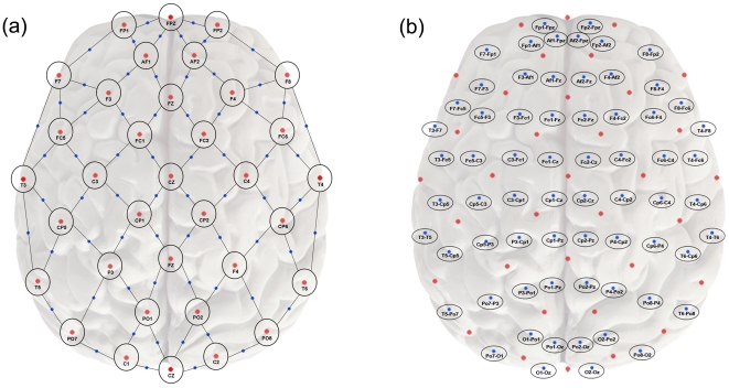
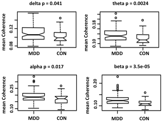
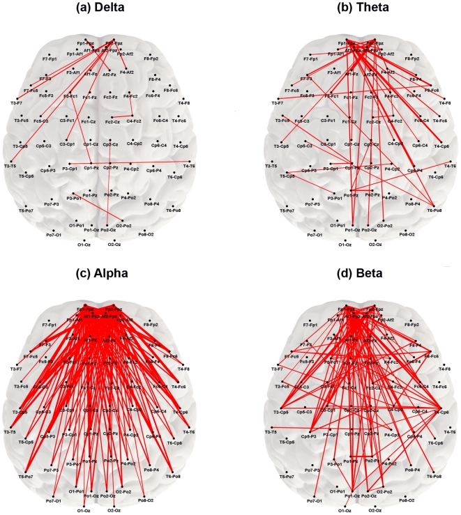
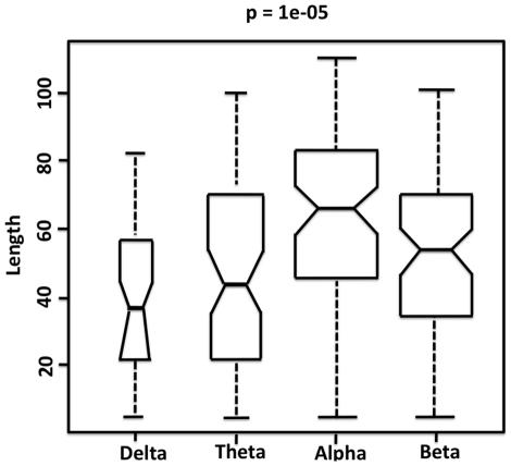
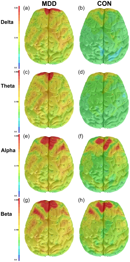
Symptoms of Major Depressive Disorder (MDD) are hypothesized to arise from dysfunction in brain networks linking the limbic system and cortical regions. Alterations in brain functional cortical connectivity in resting-state networks have been detected with functional imaging techniques, but neurophysiologic connectivity measures have not been systematically examined. We used weighted network analysis to examine resting state functional connectivity as measured by quantitative electroencephalographic (qEEG) coherence in 121 unmedicated subjects with MDD and 37 healthy controls. Subjects with MDD had significantly higher overall coherence as compared to controls in the delta (0.5-4 Hz), theta (4-8 Hz), alpha (8-12 Hz), and beta (12-20 Hz) frequency bands. The frontopolar region contained the greatest number of "hub nodes" (surface recording locations) with high connectivity. MDD subjects expressed higher theta and alpha coherence primarily in longer distance connections between frontopolar and temporal or parietooccipital regions, and higher beta coherence primarily in connections within and between electrodes overlying the dorsolateral prefrontal cortical (DLPFC) or temporal regions. Nearest centroid analysis indicated that MDD subjects were best characterized by six alpha band connections primarily involving the prefrontal region. The present findings indicate a loss of selectivity in resting functional connectivity in MDD. The overall greater coherence observed in depressed subjects establishes a new context for the interpretation of previous studies showing differences in frontal alpha power and synchrony between subjects with MDD and normal controls. These results can inform the development of qEEG state and trait biomarkers for MDD.
重度抑郁症(MDD)的症状被假设是由于连接边缘系统和皮质区域的大脑网络功能障碍引起的。在静息状态网络中,已经使用功能成像技术检测到大脑功能皮质连通性的改变,但神经生理连通性测量尚未系统地检查。我们使用加权网络分析来检查 121 名未经药物治疗的 MDD 患者和 37 名健康对照者的静息状态功能连通性,这些患者通过定量脑电图(qEEG)相干性进行测量。与对照组相比,MDD 患者在 delta(0.5-4 Hz)、theta(4-8 Hz)、alpha(8-12 Hz)和 beta(12-20 Hz)频段的整体相干性显著更高。额极区包含数量最多的高连通性“枢纽节点”(表面记录位置)。MDD 患者主要在额极区和颞区或顶枕区之间的长距离连接中表现出更高的 theta 和 alpha 相干性,而在额极区和颞区或顶枕区之间的电极上的连接中表现出更高的 beta 相干性。最近中心分析表明,MDD 患者的六个 alpha 频段连接主要涉及前额区,这些连接特征最为明显。本研究结果表明,MDD 患者静息功能连通性的选择性丧失。在抑郁患者中观察到的整体更高的相干性为解释先前研究结果提供了新的背景,这些研究结果表明 MDD 患者和正常对照组之间的额叶 alpha 功率和同步性存在差异。这些结果可以为 MDD 的 qEEG 状态和特征生物标志物的发展提供信息。