Laboratory of Neurobiology and Stem Cells, Department of Cellular Biology, University of Concepcion, Concepción, Chile.
PLoS One. 2012;7(2):e32409. doi: 10.1371/journal.pone.0032409. Epub 2012 Feb 28.
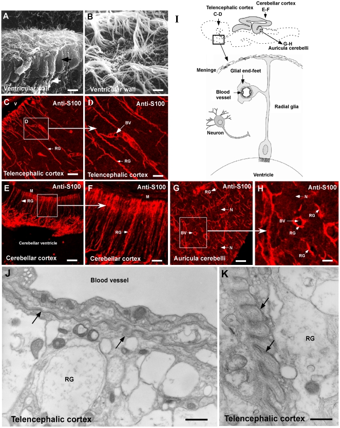
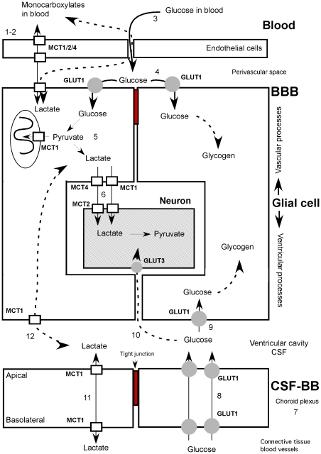
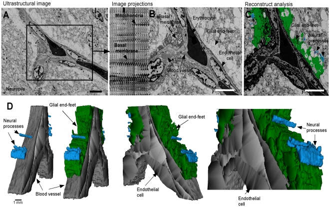
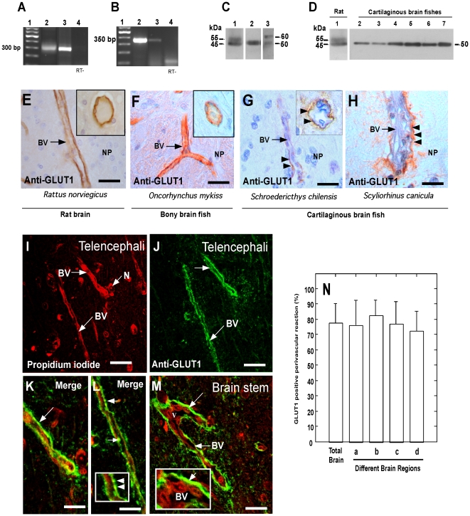
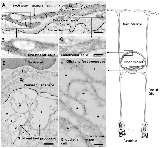
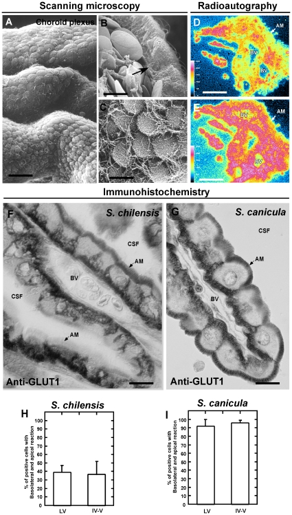
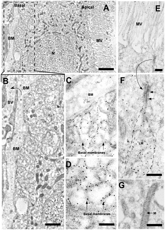
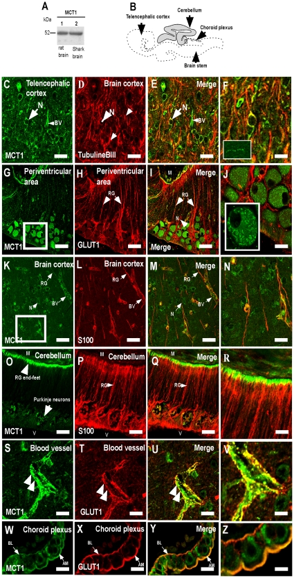
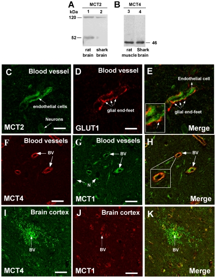
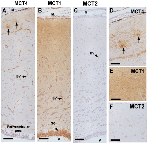
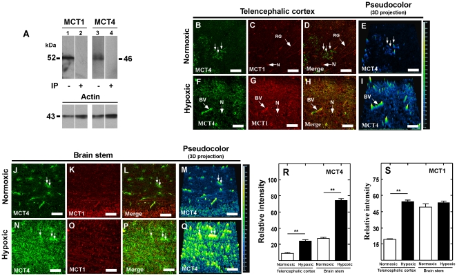
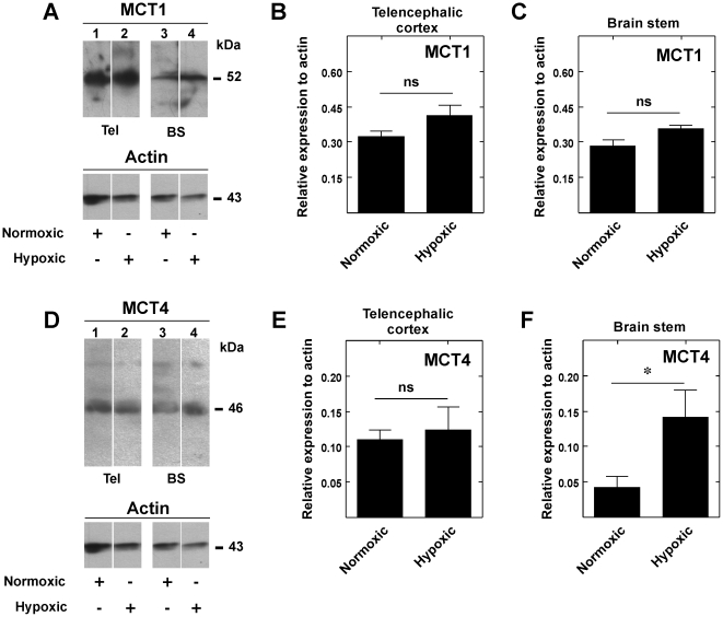
Although previous studies showed that glucose is used to support the metabolic activity of the cartilaginous fish brain, the distribution and expression levels of glucose transporter (GLUT) isoforms remained undetermined. Optic/ultrastructural immunohistochemistry approaches were used to determine the expression of GLUT1 in the glial blood-brain barrier (gBBB). GLUT1 was observed solely in glial cells; it was primarily located in end-feet processes of the gBBB. Western blot analysis showed a protein with a molecular mass of 50 kDa, and partial sequencing confirmed GLUT1 identity. Similar approaches were used to demonstrate increased GLUT1 polarization to both apical and basolateral membranes in choroid plexus epithelial cells. To explore monocarboxylate transporter (MCT) involvement in shark brain metabolism, the expression of MCTs was analyzed. MCT1, 2 and 4 were expressed in endothelial cells; however, only MCT1 and MCT4 were present in glial cells. In neurons, MCT2 was localized at the cell membrane whereas MCT1 was detected within mitochondria. Previous studies demonstrated that hypoxia modified GLUT and MCT expression in mammalian brain cells, which was mediated by the transcription factor, hypoxia inducible factor-1. Similarly, we observed that hypoxia modified MCT1 cellular distribution and MCT4 expression in shark telencephalic area and brain stem, confirming the role of these transporters in hypoxia adaptation. Finally, using three-dimensional ultrastructural microscopy, the interaction between glial end-feet and leaky blood vessels of shark brain was assessed in the present study. These data suggested that the brains of shark may take up glucose from blood using a different mechanism than that used by mammalian brains, which may induce astrocyte-neuron lactate shuttling and metabolic coupling as observed in mammalian brain. Our data suggested that the structural conditions and expression patterns of GLUT1, MCT1, MCT2 and MCT4 in shark brain may establish the molecular foundation of metabolic coupling between glia and neurons.
虽然先前的研究表明葡萄糖用于支持软骨鱼类大脑的代谢活动,但葡萄糖转运蛋白(GLUT)同工型的分布和表达水平仍未确定。采用光/超微结构免疫组织化学方法确定 GLUT1 在神经胶质血脑屏障(gBBB)中的表达。GLUT1 仅在神经胶质细胞中观察到;它主要位于 gBBB 的终足过程中。Western blot 分析显示分子量为 50 kDa 的蛋白质,部分测序证实了 GLUT1 的身份。类似的方法用于证明脉络丛上皮细胞中 GLUT1 向顶端和基底外侧膜的极化增加。为了探索单羧酸转运蛋白(MCT)在鲨鱼大脑代谢中的参与,分析了 MCT 的表达。MCT1、2 和 4 在内皮细胞中表达;然而,只有 MCT1 和 MCT4 存在于神经胶质细胞中。在神经元中,MCT2 位于细胞膜上,而 MCT1 则存在于线粒体中。先前的研究表明,缺氧改变了哺乳动物脑细胞中的 GLUT 和 MCT 表达,这是由转录因子缺氧诱导因子-1 介导的。同样,我们观察到缺氧改变了鲨鱼端脑区和脑干中 MCT1 的细胞分布和 MCT4 的表达,证实了这些转运蛋白在缺氧适应中的作用。最后,使用三维超微结构显微镜,本研究评估了鲨鱼大脑中神经胶质终足与渗漏血管之间的相互作用。这些数据表明,鲨鱼的大脑可能使用与哺乳动物大脑不同的机制从血液中摄取葡萄糖,这可能会诱导星形胶质细胞-神经元乳酸穿梭和代谢偶联,如在哺乳动物大脑中观察到的那样。我们的数据表明,鲨鱼脑中 GLUT1、MCT1、MCT2 和 MCT4 的结构条件和表达模式可能为胶质细胞和神经元之间的代谢偶联奠定分子基础。