Department of Medicine, The University of Hong Kong, Queen Mary Hospital, Hong Kong.
PLoS One. 2012;7(2):e32622. doi: 10.1371/journal.pone.0032622. Epub 2012 Feb 28.
We determined the association between various clinical parameters and significant liver injury in both hepatitis B e antigen (HBeAg)-positive and HBeAg-negative patients.
From 1994 to 2008, liver biopsy was performed on 319 treatment-naïve CHB patients. Histologic assessment was based on the Knodell histologic activity index for necroinflammation and the Ishak fibrosis staging for fibrosis.
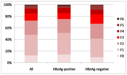
211 HBeAg-positive and 108 HBeAg-negative patients were recruited, with a median age of 31 and 46 years respectively. 9 out of 40 (22.5%) HBeAg-positive patients with normal ALT had significant histologic abnormalities (necroinflammation grading ≥ 7 or fibrosis score ≥ 3). There was a significant difference in fibrosis scores among HBeAg-positive patients with an ALT level within the Prati criteria (30 U/L for men, 19 U/L for women) and patients with a normal ALT but exceeding the Prati criteria (p = 0.024). Age, aspartate aminotransferase and platelet count were independent predictors of significant fibrosis in HBeAg-positive patients with an elevated ALT by multivariate analysis (p = 0.007, 0.047 and 0.045 respectively). HBV DNA and platelet count were predictors of significant fibrosis in HBeAg-negative disease (p = 0.020 and 0.015 respectively). An elevated ALT was not predictive of significant fibrosis for HBeAg-positive (p = 0.345) and -negative (p = 0.544) disease. There was no significant difference in fibrosis staging among ALT 1-2 × upper limit of normal (ULN) and > × 2 ULN for both HBeAg-positive (p = 0.098) and -negative (p = 0.838) disease.
An elevated ALT does not accurately predict significant liver injury. Decisions on commencing antiviral therapy should not be heavily based on a particular ALT threshold.
我们确定了乙型肝炎 e 抗原(HBeAg)阳性和 HBeAg 阴性患者中各种临床参数与显著肝损伤之间的关系。
1994 年至 2008 年,对 319 例未经治疗的慢性乙型肝炎患者进行了肝活检。组织学评估基于坏死性炎症的 Knodell 组织学活动指数和纤维化的 Ishak 分期。
纳入 211 例 HBeAg 阳性和 108 例 HBeAg 阴性患者,中位年龄分别为 31 岁和 46 岁。40 例 HBeAg 阳性 ALT 正常患者中有 9 例(22.5%)存在显著组织学异常(坏死性炎症分级≥7 或纤维化评分≥3)。在符合 Prati 标准的 ALT 水平(男性 30 U/L,女性 19 U/L)的 HBeAg 阳性患者和 ALT 正常但超过 Prati 标准的患者之间,纤维化评分存在显著差异(p=0.024)。年龄、天冬氨酸氨基转移酶和血小板计数是 HBeAg 阳性患者 ALT 升高时发生显著纤维化的独立预测因素(p=0.007、0.047 和 0.045)。HBV DNA 和血小板计数是 HBeAg 阴性疾病显著纤维化的预测因素(p=0.020 和 0.015)。升高的 ALT 对 HBeAg 阳性(p=0.345)和阴性(p=0.544)疾病的显著纤维化没有预测作用。在 HBeAg 阳性(p=0.098)和阴性(p=0.838)疾病中,ALT 为 1-2×正常值上限(ULN)和>×2ULN 之间的纤维化分期没有显著差异。
升高的 ALT 并不能准确预测显著的肝损伤。启动抗病毒治疗的决定不应过分依赖于特定的 ALT 阈值。