Shandong Provincial Hospital Affiliated to Shandong First Medical University, Jinan, Shandong, 250021, People's Republic of China.
Shandong Provincial Hospital, Cheeloo College of Medicine, Shandong University, Jinan, Shandong, 250021, People's Republic of China.
BMC Med. 2021 Oct 15;19(1):247. doi: 10.1186/s12916-021-02085-3.
We and others have confirmed activation of macrophages plays a critical role in liver injury and fibrogenesis during HBV infection. And we have also proved HBeAg can obviously induce the production of macrophage inflammatory cytokines compared with HBsAg and HBcAg. However, the receptor and functional domain of HBeAg in macrophage activation and its effects and mechanisms on hepatic fibrosis remain elusive.
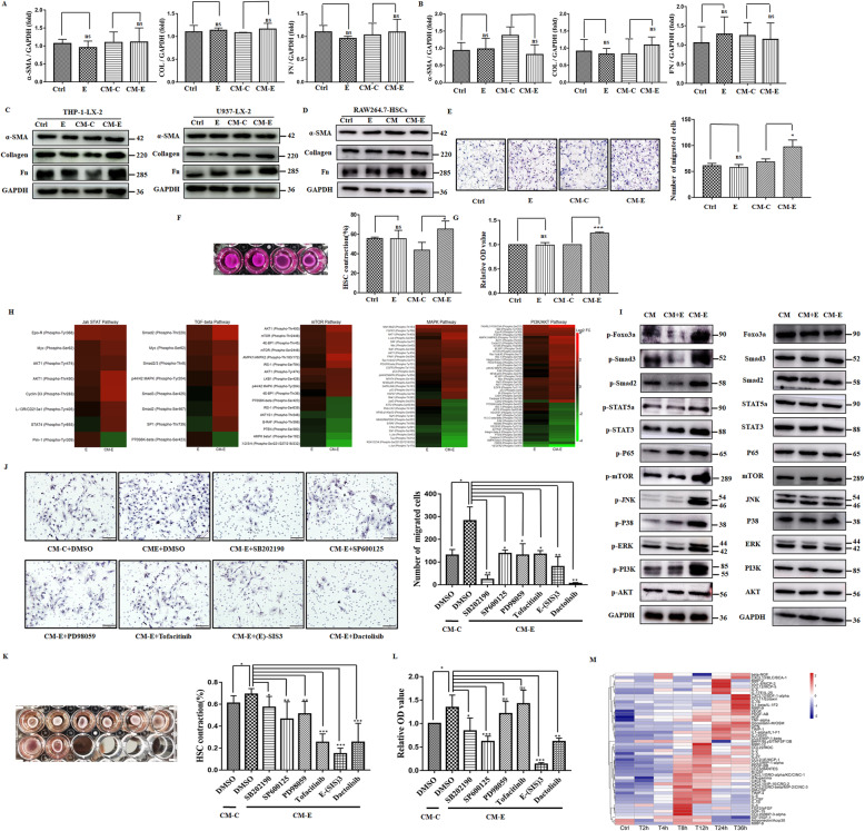
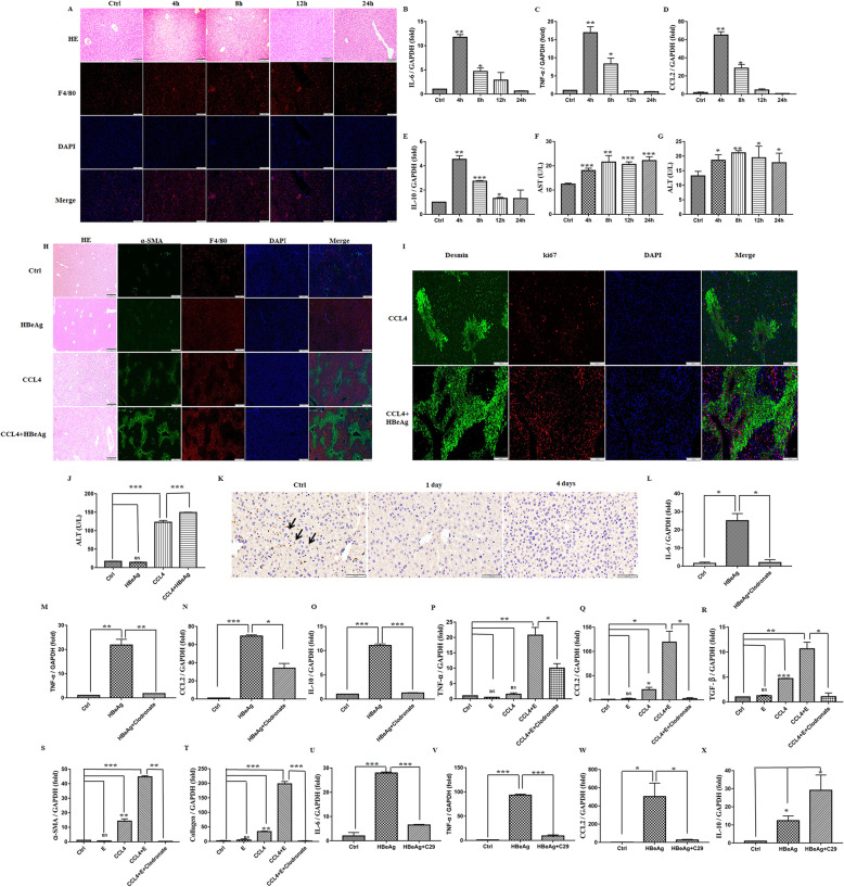
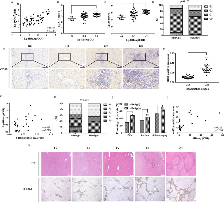
The potentially direct binding receptors of HBeAg were screened and verified by Co-IP assay. Meanwhile, the function domain and accessible peptides of HBeAg for macrophage activation were analyzed by prediction of surface accessible peptide, construction, and synthesis of truncated fragments. Furthermore, effects and mechanisms of the activation of hepatic stellate cells induced by HBeAg-treated macrophages were investigated by Transwell, CCK-8, Gel contraction assay, Phospho Explorer antibody microarray, and Luminex assay. Finally, the effect of HBeAg in hepatic inflammation and fibrosis was evaluated in both human and murine tissues by immunohistochemistry, immunofluorescence, ELISA, and detection of liver enzymes.
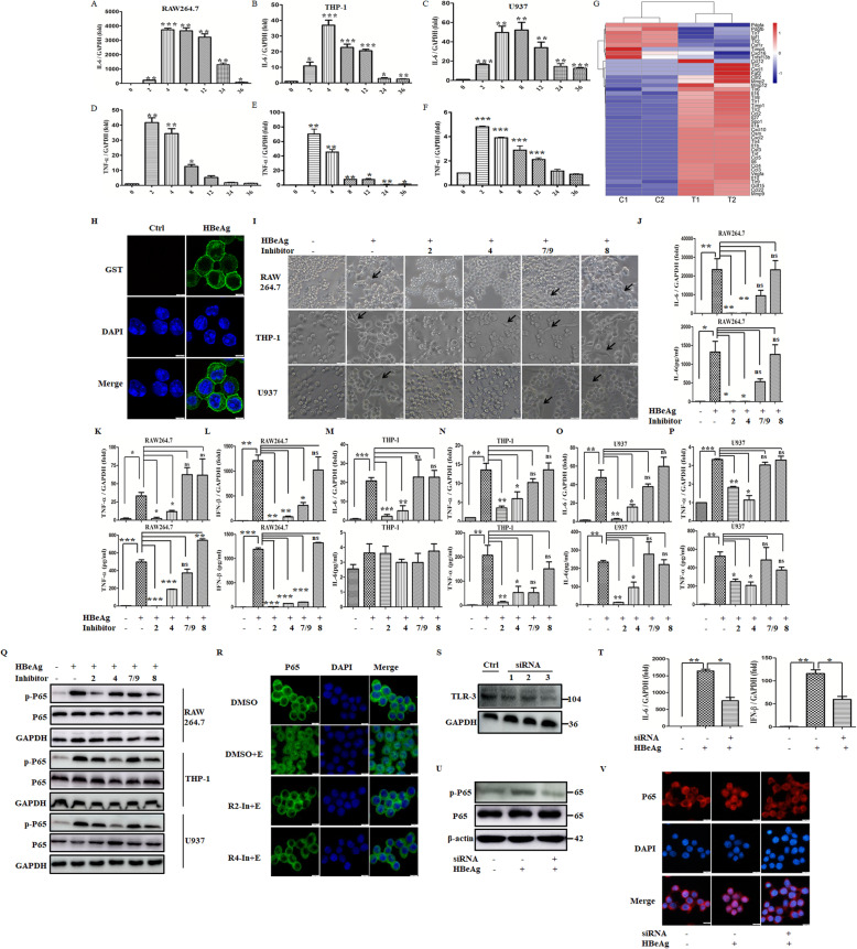
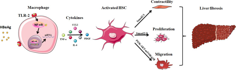
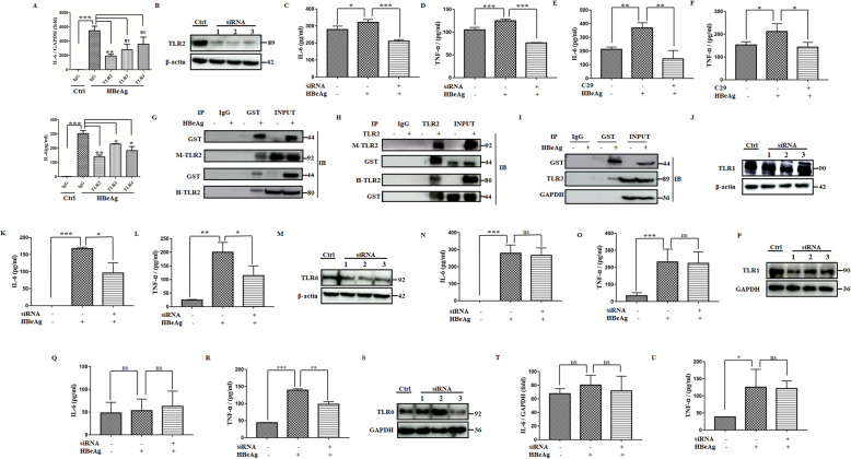
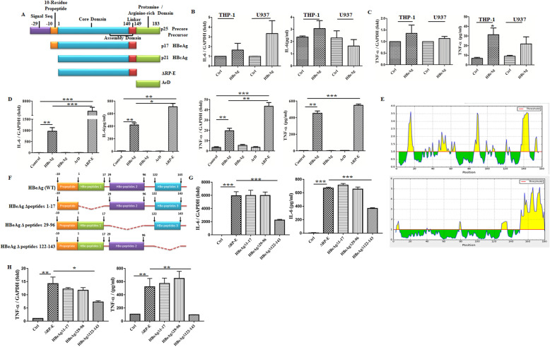
Herein, we verified TLR-2 was the direct binding receptor of HBeAg. Meanwhile, C-terminal peptide (122-143 aa.) of core domain in HBeAg was critical for macrophage activation. But arginine-rich domain of HBcAg hided this function, although HBcAg and HBeAg shared the same core domain. Furthermore, HBeAg promoted the proliferation, motility, and contraction of hepatic stellate cells (HSCs) in a macrophage-dependent manner, but not alone. PI3K-AKT-mTOR and p38 MAPK signaling pathway were responsible for motility phenotype of HSCs, while the Smad-dependent TGF-β signaling pathway for proliferation and contraction of them. Additionally, multiple chemokines and cytokines, such as CCL2, CCL5, CXCL10, and TNF-α, might be key mediators of HSC activation. Consistently, HBeAg induced transient inflammation response and promoted early fibrogenesis via TLR-2 in mice. Finally, clinical investigations suggested that the level of HBeAg is associated with inflammation and fibrosis degrees in patients infected with HBV.
HBeAg activated macrophages via the TLR-2/NF-κB signal pathway and further exacerbated hepatic fibrosis by facilitating motility, proliferation, and contraction of HSCs with the help of macrophages.
我们和其他人已经证实,HBV 感染期间,巨噬细胞的激活在肝损伤和纤维化中起着关键作用。我们还证明,与 HBsAg 和 HBcAg 相比,HBeAg 可以明显诱导巨噬细胞产生炎症细胞因子。然而,HBeAg 在巨噬细胞激活中的受体和功能域及其对肝纤维化的影响和机制仍不清楚。
通过 Co-IP 测定筛选和验证 HBeAg 的潜在直接结合受体。同时,通过预测表面可及肽、构建和合成截短片段分析 HBeAg 用于巨噬细胞激活的功能域和可及肽。此外,通过 Transwell、CCK-8、凝胶收缩测定、Phospho Explorer 抗体微阵列和 Luminex 测定研究 HBeAg 处理的巨噬细胞诱导的肝星状细胞的激活作用及其机制。最后,通过免疫组化、免疫荧光、ELISA 和检测肝酶在人和鼠组织中评估 HBeAg 在肝炎症和纤维化中的作用。
在此,我们验证了 TLR-2 是 HBeAg 的直接结合受体。同时,HBeAg 核心域的 C 端肽(122-143aa.)对于巨噬细胞激活是关键的。但是 HBcAg 的富含精氨酸的结构域隐藏了这种功能,尽管 HBcAg 和 HBeAg 共享相同的核心域。此外,HBeAg 以巨噬细胞依赖的方式促进肝星状细胞(HSCs)的增殖、迁移和收缩,但不是单独作用。PI3K-AKT-mTOR 和 p38 MAPK 信号通路负责 HSCs 的迁移表型,而 Smad 依赖性 TGF-β信号通路负责其增殖和收缩。此外,多种趋化因子和细胞因子,如 CCL2、CCL5、CXCL10 和 TNF-α,可能是 HSC 激活的关键介质。一致地,HBeAg 通过 TLR-2 在小鼠中诱导短暂的炎症反应,并通过促进增殖和收缩来促进早期纤维化。最后,临床研究表明,HBV 感染患者的 HBeAg 水平与炎症和纤维化程度相关。
HBeAg 通过 TLR-2/NF-κB 信号通路激活巨噬细胞,并通过巨噬细胞促进 HSCs 的迁移、增殖和收缩,进一步加剧肝纤维化。