• 文献检索
  • 文档翻译
  • 深度研究
  • 学术资讯
  • Suppr Zotero 插件Zotero 插件
  • 邀请有礼
  • 套餐&价格
  • 历史记录
应用&插件
Suppr Zotero 插件Zotero 插件浏览器插件Mac 客户端Windows 客户端微信小程序
定价
高级版会员购买积分包购买API积分包
服务
文献检索文档翻译深度研究API 文档MCP 服务
关于我们
关于 Suppr公司介绍联系我们用户协议隐私条款
关注我们

Suppr 超能文献

核心技术专利:CN118964589B侵权必究
粤ICP备2023148730 号-1Suppr @ 2026

文献检索

告别复杂PubMed语法,用中文像聊天一样搜索,搜遍4000万医学文献。AI智能推荐,让科研检索更轻松。

立即免费搜索

文件翻译

保留排版,准确专业,支持PDF/Word/PPT等文件格式,支持 12+语言互译。

免费翻译文档

深度研究

AI帮你快速写综述,25分钟生成高质量综述,智能提取关键信息,辅助科研写作。

立即免费体验

氧化铁掺入对三种人源细胞类型的软骨生成潜力的影响。

The effects of iron oxide incorporation on the chondrogenic potential of three human cell types.

机构信息

Biomaterials and Tissue Engineering Group, Leeds Dental Institute, University of Leeds, UK.

出版信息

J Tissue Eng Regen Med. 2013 Jun;7(6):461-9. doi: 10.1002/term.544. Epub 2012 Mar 7.

DOI:10.1002/term.544
PMID:22396122
原文链接:https://pmc.ncbi.nlm.nih.gov/articles/PMC3747461/
Abstract

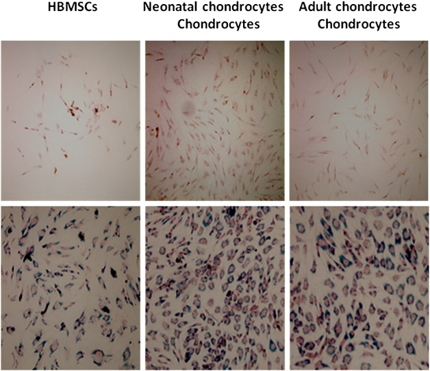
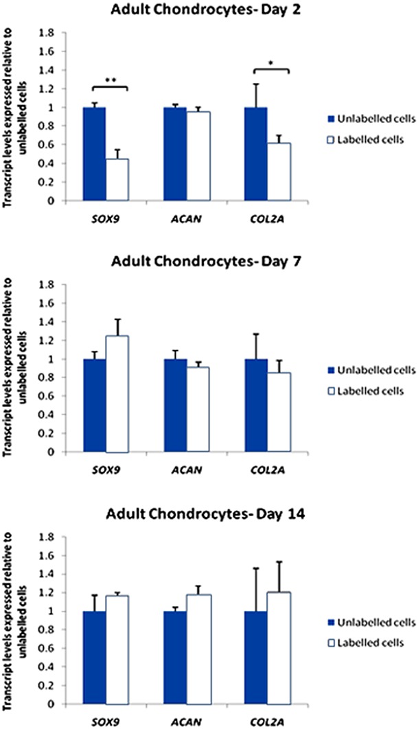
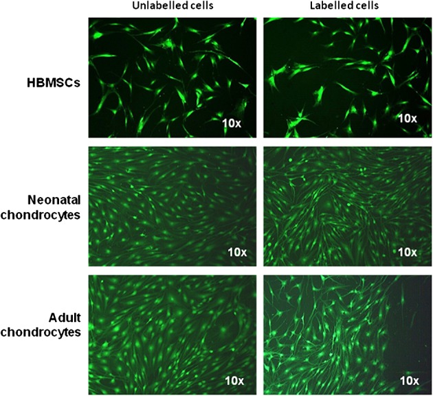
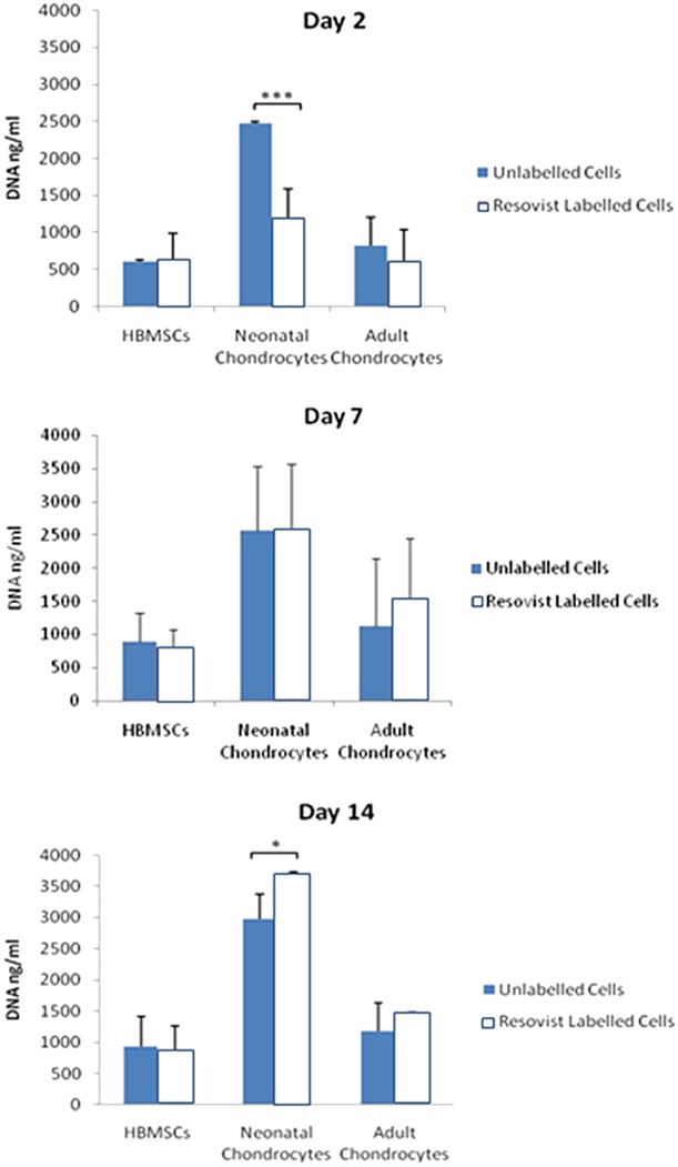
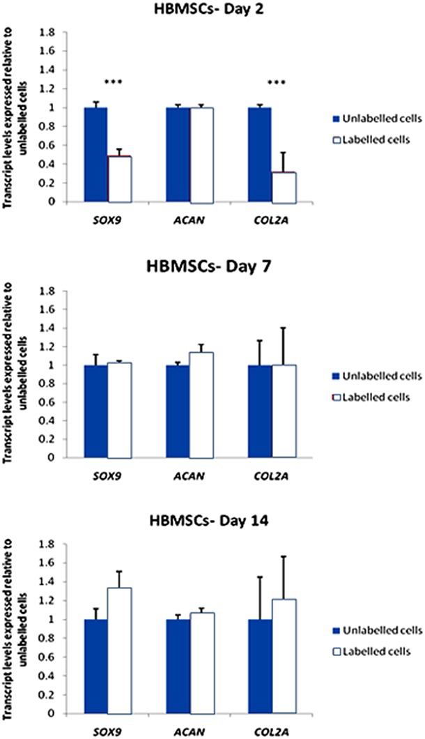
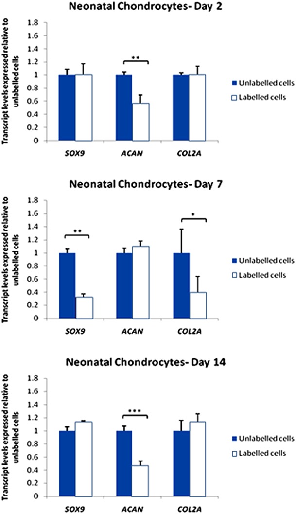
Non-invasive monitoring of living cells in vivo provides an important tool in the development of cell-based therapies in cartilage tissue engineering. High-resolution magnetic resonance imaging (MRI) has been used to monitor target cell populations in vivo. However, the side-effects on cell function of the labelling reagents, such as superparamagnetic iron oxide (SPIO), are still unclear. This study investigated the effect of SPIO particles on the chondrogenic differentiation of human bone marrow stromal cells (HBMSCs), neonatal and adult chondrocytes in vitro. Cells were labelled with SPIO for 24 h and chondrogenesis induced in serum-free medium including TGFβ3. For labelled/unlabelled cells, viability, morphology and proliferation were determined using CellTracker™ Green and PicoGreen dsDNA assays. The expression of SOX9, COL2A1 and ACAN was investigated using qRT-PCR after 2, 7 and 14 days. The results showed that viability was unaffected in all of the cells but cell morphology changed towards a 'stretched' phenotype following SPIO uptake. Cell proliferation was reduced only for labelled neonatal chondrocytes. SOX9 and COL2A1 expression decreased at day 2 but not at days 7 and 14 for labelled HBMSCs and adult chondrocytes; ACAN expression was unaffected. In contrast, SOX9 and COL2A1 expression were unaffected in labelled neonatal chondrocytes but a decrease in ACAN expression was seen at day 14. The results suggest that downregulation of chondrogenic genes associated with SPIO labelling is temporary and target cell-dependent. Resovist® can be used to label HBMSCs or mature chondrocytes for MR imaging of cells for cartilage tissue engineering.

摘要

在软骨组织工程中,对活细胞进行非侵入性监测为基于细胞的治疗方法的发展提供了重要工具。高分辨率磁共振成像(MRI)已被用于监测体内的靶细胞群体。然而,标记试剂(如超顺磁氧化铁(SPIO))对细胞功能的副作用尚不清楚。本研究调查了 SPIO 颗粒对体外人骨髓基质细胞(HBMSCs)、新生儿和成年软骨细胞的软骨分化的影响。细胞用 SPIO 标记 24 小时,并在包含 TGFβ3 的无血清培养基中诱导软骨生成。对于标记/未标记的细胞,使用 CellTracker™ Green 和 PicoGreen dsDNA 测定法测定细胞活力、形态和增殖。使用 qRT-PCR 在第 2、7 和 14 天检测 SOX9、COL2A1 和 ACAN 的表达。结果表明,所有细胞的活力均未受影响,但 SPIO 摄取后细胞形态向“伸展”表型改变。仅标记的新生儿软骨细胞的细胞增殖减少。标记的 HBMSCs 和成年软骨细胞的 SOX9 和 COL2A1 表达在第 2 天下降,但在第 7 和 14 天没有下降;ACAN 表达不受影响。相比之下,标记的新生儿软骨细胞的 SOX9 和 COL2A1 表达不受影响,但在第 14 天观察到 ACAN 表达下降。结果表明,与 SPIO 标记相关的软骨生成基因的下调是暂时的且与靶细胞有关。Resovist®可用于标记 HBMSCs 或成熟软骨细胞,以进行软骨组织工程中细胞的 MRI 成像。

https://cdn.ncbi.nlm.nih.gov/pmc/blobs/9917/3747461/57d4ca5faccf/term0007-0461-f6.jpg
https://cdn.ncbi.nlm.nih.gov/pmc/blobs/9917/3747461/b6b2ac510728/term0007-0461-f1.jpg
https://cdn.ncbi.nlm.nih.gov/pmc/blobs/9917/3747461/4a3b9d5f86f2/term0007-0461-f2.jpg
https://cdn.ncbi.nlm.nih.gov/pmc/blobs/9917/3747461/cf8e79125d44/term0007-0461-f3.jpg
https://cdn.ncbi.nlm.nih.gov/pmc/blobs/9917/3747461/29fbad66b301/term0007-0461-f4.jpg
https://cdn.ncbi.nlm.nih.gov/pmc/blobs/9917/3747461/ba81b3f560db/term0007-0461-f5.jpg
https://cdn.ncbi.nlm.nih.gov/pmc/blobs/9917/3747461/57d4ca5faccf/term0007-0461-f6.jpg

相似文献

1
The effects of iron oxide incorporation on the chondrogenic potential of three human cell types.氧化铁掺入对三种人源细胞类型的软骨生成潜力的影响。
J Tissue Eng Regen Med. 2013 Jun;7(6):461-9. doi: 10.1002/term.544. Epub 2012 Mar 7.
2
Dose-response of superparamagnetic iron oxide labeling on mesenchymal stem cells chondrogenic differentiation: a multi-scale in vitro study.超顺磁性氧化铁标记对间充质干细胞软骨分化的剂量反应:多尺度体外研究。
PLoS One. 2014 May 30;9(5):e98451. doi: 10.1371/journal.pone.0098451. eCollection 2014.
3
Cell viability and chondrogenic differentiation capability of human mesenchymal stem cells after iron labeling with iron sucrose.用蔗糖铁标记后人骨髓间充质干细胞的细胞活力和软骨分化能力
Stem Cells Dev. 2014 Nov 1;23(21):2568-80. doi: 10.1089/scd.2014.0153. Epub 2014 Aug 25.
4
Clinically translatable cell tracking and quantification by MRI in cartilage repair using superparamagnetic iron oxides.临床可转化的细胞示踪和定量磁共振成像在软骨修复中使用超顺磁性氧化铁。
PLoS One. 2011 Feb 23;6(2):e17001. doi: 10.1371/journal.pone.0017001.
5
Comparison of incremental concentrations of micron-sized superparamagnetic iron oxide for labelling articular cartilage derived chondroprogenitors.比较微尺度超顺磁氧化铁的递增浓度标记关节软骨来源的软骨祖细胞。
Acta Histochem. 2019 Oct;121(7):791-797. doi: 10.1016/j.acthis.2019.07.004. Epub 2019 Jul 18.
6
[Effect of superparamagnetic iron oxide on differentiation of rat bone marrow stem cells into chondrocytes in vitro].超顺磁性氧化铁对大鼠骨髓干细胞体外向软骨细胞分化的影响
Nan Fang Yi Ke Da Xue Xue Bao. 2017 May 20;37(5):652-658. doi: 10.3969/j.issn.1673-4254.2017.05.14.
7
Direct labeling of hMSC with SPIO: the long-term influence on toxicity, chondrogenic differentiation capacity, and intracellular distribution.SPIO 直接标记 hMSC:对毒性、软骨分化能力和细胞内分布的长期影响。
Mol Imaging Biol. 2011 Jun;13(3):443-451. doi: 10.1007/s11307-010-0360-7.
8
Magnetic resonance imaging of umbilical cord stem cells labeled with superparamagnetic iron oxide nanoparticles: effects of labelling and transplantation parameters.超顺磁氧化铁纳米颗粒标记的脐带干细胞的磁共振成像:标记和移植参数的影响。
Sci Rep. 2020 Aug 13;10(1):13684. doi: 10.1038/s41598-020-70291-9.
9
MicroRNA regulation associated chondrogenesis of mouse MSCs grown on polyhydroxyalkanoates.聚羟基烷酸酯上培养的小鼠间充质干细胞的 microRNA 调控与软骨生成相关。
Biomaterials. 2011 Sep;32(27):6435-44. doi: 10.1016/j.biomaterials.2011.05.031. Epub 2011 Jun 12.
10
Comparative study of the chondrogenic potential of human bone marrow stromal cells, neonatal chondrocytes and adult chondrocytes.人骨髓基质细胞、新生儿软骨细胞和成人软骨细胞的成软骨潜力比较研究。
Biochem Biophys Res Commun. 2010 Oct 22;401(3):333-8. doi: 10.1016/j.bbrc.2010.09.042. Epub 2010 Sep 16.

引用本文的文献

1
The Role of Cartilage Stem/Progenitor Cells in Cartilage Repair in Osteoarthritis.软骨干细胞/前体细胞在骨关节炎软骨修复中的作用。
Curr Stem Cell Res Ther. 2023;18(7):892-903. doi: 10.2174/1574888X17666221006113739.
2
Iron Oxide Nanoparticles in Regenerative Medicine and Tissue Engineering.再生医学与组织工程中的氧化铁纳米颗粒
Nanomaterials (Basel). 2021 Sep 8;11(9):2337. doi: 10.3390/nano11092337.
3
Magnetic resonance imaging of umbilical cord stem cells labeled with superparamagnetic iron oxide nanoparticles: effects of labelling and transplantation parameters.

本文引用的文献

1
Comparative study of the chondrogenic potential of human bone marrow stromal cells, neonatal chondrocytes and adult chondrocytes.人骨髓基质细胞、新生儿软骨细胞和成人软骨细胞的成软骨潜力比较研究。
Biochem Biophys Res Commun. 2010 Oct 22;401(3):333-8. doi: 10.1016/j.bbrc.2010.09.042. Epub 2010 Sep 16.
2
Labeling of human mesenchymal stromal cells with superparamagnetic iron oxide leads to a decrease in migration capacity and colony formation ability.用超顺磁性氧化铁标记人间充质基质细胞会导致其迁移能力和集落形成能力下降。
Cytotherapy. 2009;11(1):68-78. doi: 10.1080/14653240802666043.
3
Effects of iron oxide incorporation for long term cell tracking on MSC differentiation in vitro and in vivo.
超顺磁氧化铁纳米颗粒标记的脐带干细胞的磁共振成像:标记和移植参数的影响。
Sci Rep. 2020 Aug 13;10(1):13684. doi: 10.1038/s41598-020-70291-9.
4
Iron oxide nanoparticles promote the migration of mesenchymal stem cells to injury sites.氧化铁纳米颗粒促进间充质干细胞向损伤部位迁移。
Int J Nanomedicine. 2019 Jan 14;14:573-589. doi: 10.2147/IJN.S184920. eCollection 2019.
5
Stem cells as delivery vehicles for regenerative medicine-challenges and perspectives.干细胞作为再生医学的载体——挑战与展望
World J Stem Cells. 2018 May 26;10(5):43-56. doi: 10.4252/wjsc.v10.i5.43.
6
Non-invasive monitoring of hydrogel degradation and cartilage regeneration by multiparametric MR imaging.多参数磁共振成像无创监测水凝胶降解及软骨再生
Theranostics. 2018 Jan 13;8(4):1146-1158. doi: 10.7150/thno.22514. eCollection 2018.
7
[Effect of superparamagnetic iron oxide on differentiation of rat bone marrow stem cells into chondrocytes in vitro].超顺磁性氧化铁对大鼠骨髓干细胞体外向软骨细胞分化的影响
Nan Fang Yi Ke Da Xue Xue Bao. 2017 May 20;37(5):652-658. doi: 10.3969/j.issn.1673-4254.2017.05.14.
8
Superparamagnetic iron oxide nanoparticle targeting of adipose tissue-derived stem cells in diabetes-associated erectile dysfunction.超顺磁性氧化铁纳米颗粒靶向脂肪组织来源干细胞治疗糖尿病相关性勃起功能障碍
Asian J Androl. 2017 Jul-Aug;19(4):425-432. doi: 10.4103/1008-682X.179532.
9
Cellular magnetic resonance imaging contrast generated by the ferritin heavy chain genetic reporter under the control of a Tet-On switch.在Tet-On开关控制下,由铁蛋白重链基因报告基因产生的细胞磁共振成像对比。
Stem Cell Res Ther. 2015 Oct 31;6:207. doi: 10.1186/s13287-015-0205-z.
10
Dose-response of superparamagnetic iron oxide labeling on mesenchymal stem cells chondrogenic differentiation: a multi-scale in vitro study.超顺磁性氧化铁标记对间充质干细胞软骨分化的剂量反应:多尺度体外研究。
PLoS One. 2014 May 30;9(5):e98451. doi: 10.1371/journal.pone.0098451. eCollection 2014.
长期细胞追踪中掺入氧化铁对间充质干细胞体外和体内分化的影响。
Biochem Biophys Res Commun. 2008 May 16;369(4):1076-81. doi: 10.1016/j.bbrc.2008.02.159. Epub 2008 Mar 10.
4
Carboxylated superparamagnetic iron oxide particles label cells intracellularly without transfection agents.羧化超顺磁性氧化铁颗粒无需转染剂即可在细胞内标记细胞。
Mol Imaging Biol. 2008 May-Jun;10(3):138-46. doi: 10.1007/s11307-007-0130-3. Epub 2008 Feb 23.
5
Development of custom-built bone scaffolds using mesenchymal stem cells and apatite-wollastonite glass-ceramics.利用间充质干细胞和磷灰石-硅灰石微晶玻璃开发定制骨支架。
Tissue Eng. 2007 Dec;13(12):2891-901. doi: 10.1089/ten.2007.0124.
6
Differential regulation of lubricin/superficial zone protein by transforming growth factor beta/bone morphogenetic protein superfamily members in articular chondrocytes and synoviocytes.转化生长因子β/骨形态发生蛋白超家族成员对关节软骨细胞和滑膜细胞中润滑素/表面区蛋白的差异调节
Arthritis Rheum. 2007 Jul;56(7):2312-21. doi: 10.1002/art.22659.
7
In vivo magnetic resonance imaging of iron oxide-labeled, arterially-injected mesenchymal stem cells in kidneys of rats with acute ischemic kidney injury: detection and monitoring at 3T.急性缺血性肾损伤大鼠肾脏中经动脉注射的氧化铁标记间充质干细胞的体内磁共振成像:3T 下的检测与监测
J Magn Reson Imaging. 2007 Jun;25(6):1179-91. doi: 10.1002/jmri.20925.
8
A comparison between the chondrogenic potential of human bone marrow stem cells (BMSCs) and adipose-derived stem cells (ADSCs) taken from the same donors.对取自相同供体的人骨髓干细胞(BMSCs)和脂肪来源干细胞(ADSCs)的软骨形成潜能进行比较。
Tissue Eng. 2007 Apr;13(4):659-66. doi: 10.1089/ten.2006.0118.
9
Identification of markers to characterize and sort human articular chondrocytes with enhanced in vitro chondrogenic capacity.鉴定用于表征和分选具有增强体外软骨形成能力的人关节软骨细胞的标志物。
Arthritis Rheum. 2007 Feb;56(2):586-95. doi: 10.1002/art.22408.
10
Molecular imaging using visible light to reveal biological changes in the brain.利用可见光进行分子成像以揭示大脑中的生物学变化。
Neuroimaging Clin N Am. 2006 Nov;16(4):633-54, ix. doi: 10.1016/j.nic.2006.08.002.