Department of Cell Biology, Miller School of Medicine, University of Miami, Miami, FL 33136, USA.
Mol Biol Cell. 2012 May;23(9):1664-74. doi: 10.1091/mbc.E11-12-0988. Epub 2012 Mar 7.
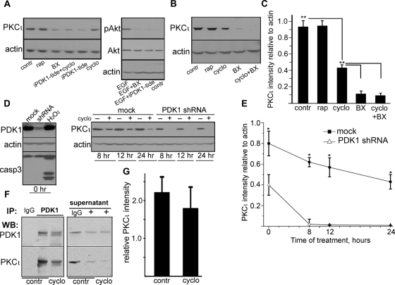
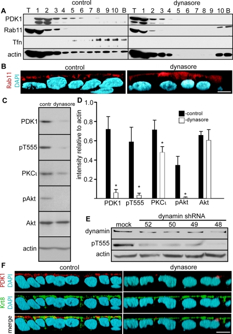
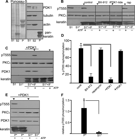
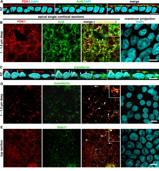
Phosphorylation of the activation domain of protein kinase C (PKC) isoforms is essential to start a conformational change that results in an active catalytic domain. This activation is necessary not only for newly synthesized molecules, but also for kinase molecules that become dephosphorylated and need to be refolded and rephosphorylated. This "rescue" mechanism is responsible for the maintenance of the steady-state levels of atypical PKC (aPKC [PKCι/λ and ζ]) and is blocked in inflammation. Although there is consensus that phosphoinositide-dependent protein kinase 1 (PDK1) is the activating kinase for newly synthesized molecules, it is unclear what kinase performs that function during the rescue and where the rescue takes place. To identify the activating kinase during the rescue mechanism, we inhibited protein synthesis and analyzed the stability of the remaining aPKC pool. PDK1 knockdown and two different PDK1 inhibitors-BX-912 and a specific pseudosubstrate peptide-destabilized PKCι. PDK1 coimmunoprecipitated with PKCι in cells without protein synthesis, confirming that the interaction is direct. In addition, we showed that PDK1 aids the rescue of aPKC in in vitro rephosphorylation assays using immunodepletion and rescue with recombinant protein. Surprisingly, we found that in Caco-2 epithelial cells and intestinal crypt enterocytes PDK1 distributes to an apical membrane compartment comprising plasma membrane and apical endosomes, which, in turn, are in close contact with intermediate filaments. PDK1 comigrated with the Rab11 compartment and, to some extent, with the transferrin compartment in sucrose gradients. PDK1, pT555-aPKC, and pAkt were dependent on dynamin activity. These results highlight a novel signaling function of apical endosomes in polarized cells.
蛋白激酶 C(PKC)同工型激活结构域的磷酸化对于启动构象变化从而产生活性催化结构域至关重要。这种激活不仅对于新合成的分子是必需的,而且对于需要去磷酸化并重新折叠和再磷酸化的激酶分子也是必需的。这种“挽救”机制负责维持非典型 PKC(aPKC [PKCι/λ 和 ζ])的稳态水平,并在炎症中被阻断。尽管人们普遍认为磷脂依赖性蛋白激酶 1(PDK1)是新合成分子的激活激酶,但尚不清楚在挽救过程中哪种激酶发挥该功能,以及挽救发生在哪里。为了在挽救机制中鉴定激活激酶,我们抑制了蛋白质合成并分析了剩余的 aPKC 池的稳定性。PDK1 敲低和两种不同的 PDK1 抑制剂-BX-912 和特定的伪底物肽-使 PKCι 不稳定。PDK1 在没有蛋白质合成的细胞中与 PKCι 共免疫沉淀,证实了这种相互作用是直接的。此外,我们还表明,PDK1 在使用免疫沉淀和重组蛋白挽救的体外再磷酸化测定中有助于 aPKC 的挽救。令人惊讶的是,我们发现在 Caco-2 上皮细胞和肠隐窝肠上皮细胞中,PDK1 分布在一个顶端膜隔室中,该隔室包括质膜和顶端内体,而顶端内体又与中间丝紧密接触。PDK1 与 Rab11 隔室共迁移,在一定程度上与蔗糖梯度中的转铁蛋白隔室共迁移。PDK1、pT555-aPKC 和 pAkt 依赖于 dynamin 活性。这些结果突出了顶端内体在极化细胞中的一种新的信号转导功能。