Department of Biomedica l Engineering, University of Wisconsin-Madison, Madison, Wisconsin, USA.
Nat Protoc. 2012 Mar 8;7(4):654-69. doi: 10.1038/nprot.2012.009.
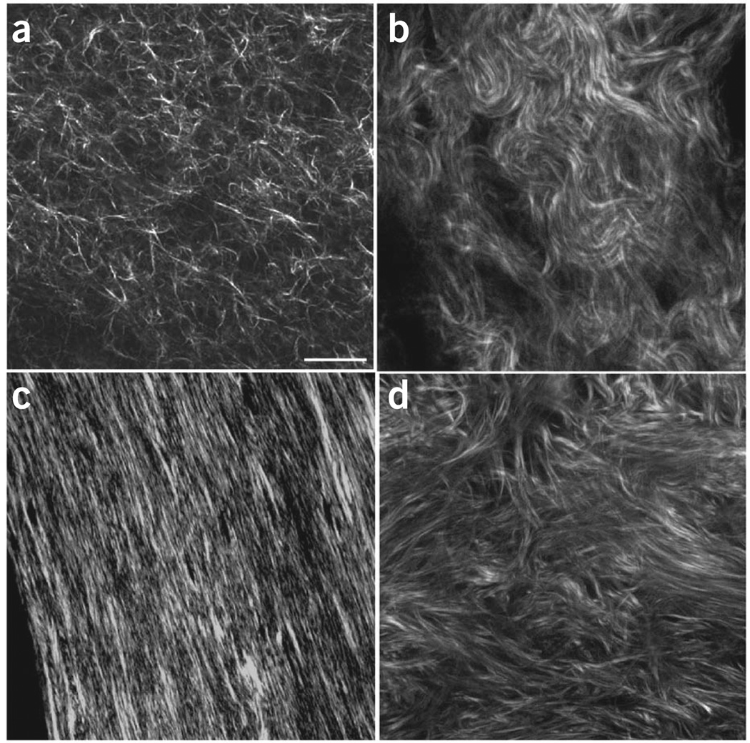
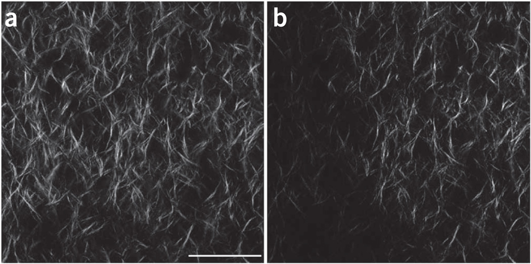
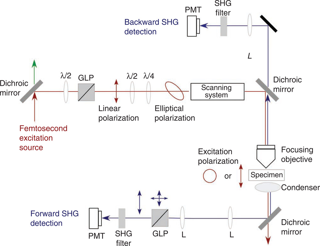
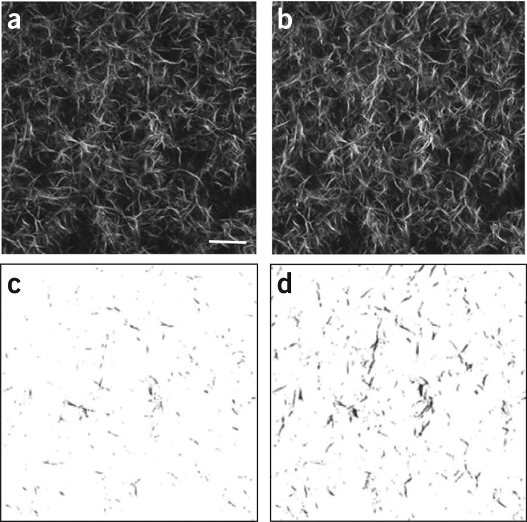
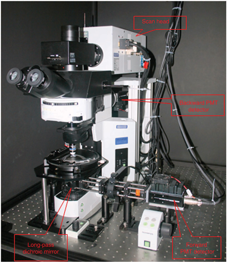
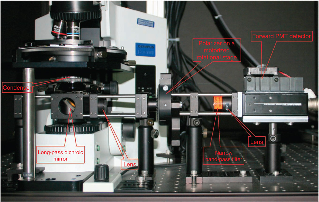
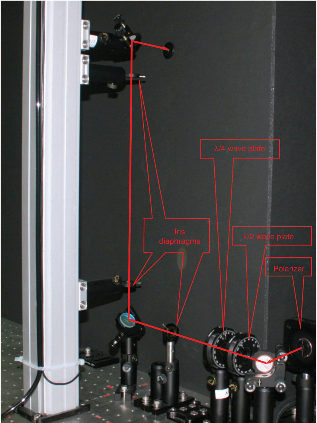
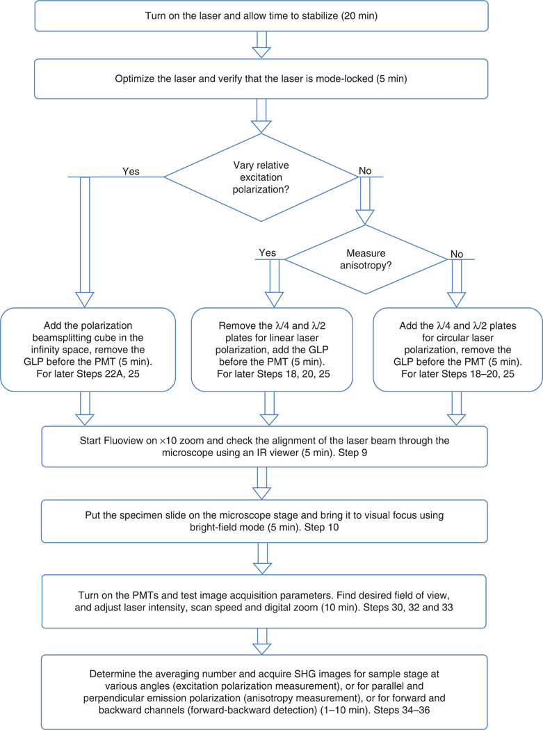
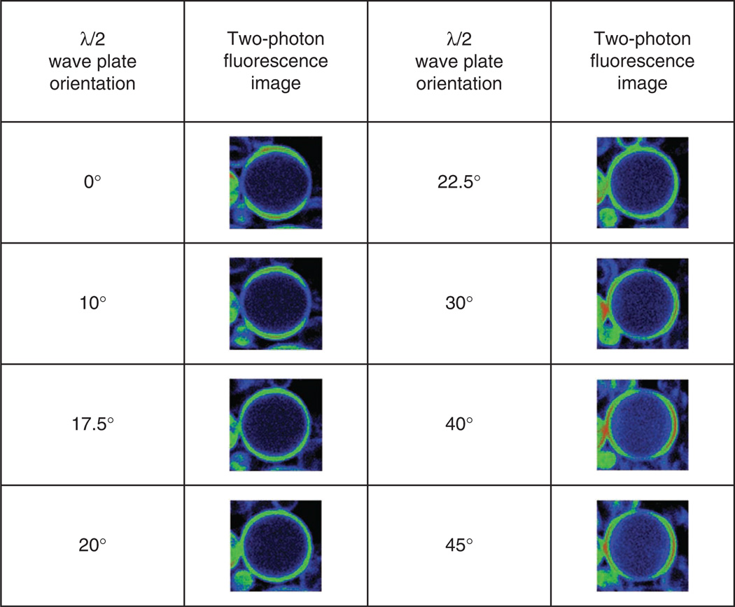
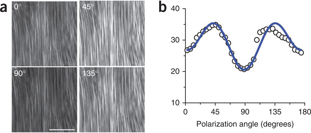
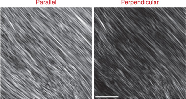
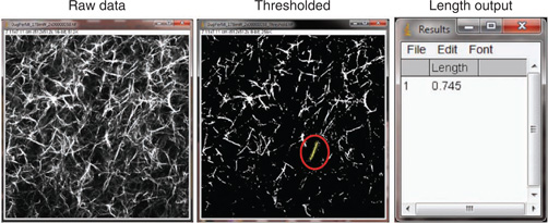
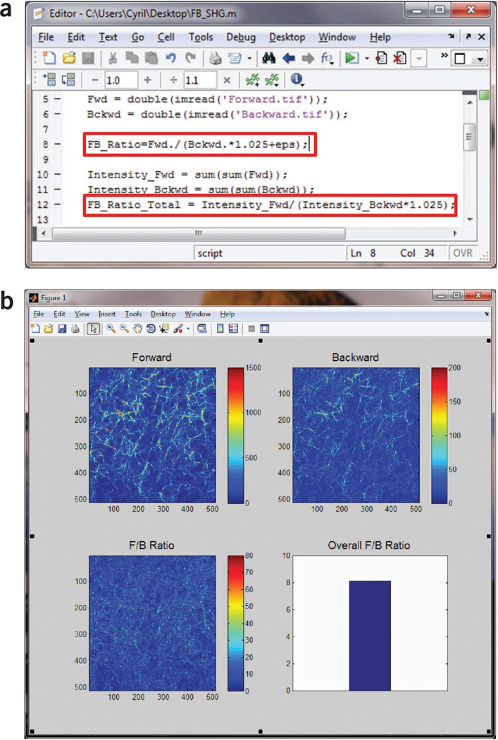
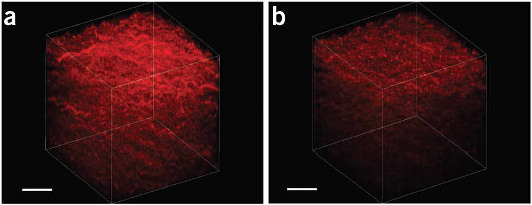
Second-harmonic generation (SHG) microscopy has emerged as a powerful modality for imaging fibrillar collagen in a diverse range of tissues. Because of its underlying physical origin, it is highly sensitive to the collagen fibril/fiber structure, and, importantly, to changes that occur in diseases such as cancer, fibrosis and connective tissue disorders. We discuss how SHG can be used to obtain more structural information on the assembly of collagen in tissues than is possible by other microscopy techniques. We first provide an overview of the state of the art and the physical background of SHG microscopy, and then describe the optical modifications that need to be made to a laser-scanning microscope to enable the measurements. Crucial aspects for biomedical applications are the capabilities and limitations of the different experimental configurations. We estimate that the setup and calibration of the SHG instrument from its component parts will require 2-4 weeks, depending on the level of the user's experience.
二次谐波产生(SHG)显微镜已成为一种强大的成像技术,可用于对各种组织中的纤维状胶原蛋白进行成像。由于其潜在的物理起源,它对胶原蛋白纤维/纤维结构非常敏感,而且重要的是,对癌症、纤维化和结缔组织疾病等疾病发生的变化非常敏感。我们将讨论 SHG 如何用于从组织结构上获得比其他显微镜技术更多的关于胶原蛋白在组织中组装的信息。我们首先提供 SHG 显微镜技术的最新发展和物理背景的概述,然后描述需要对激光扫描显微镜进行的光学修改,以实现这些测量。对于生物医学应用来说,至关重要的是不同实验配置的功能和局限性。我们估计,从组件部分搭建和校准 SHG 仪器需要 2-4 周的时间,具体取决于用户经验水平。