Miguchi Masashi, Iseki Masahiko, Shimatani Kunihiko
Department of Surgery, National Hospital Organization Hiroshimanishi Medical Center, Otake City, Japan.
Case Rep Gastroenterol. 2012 Jan;6(1):52-7. doi: 10.1159/000336320. Epub 2012 Jan 25.
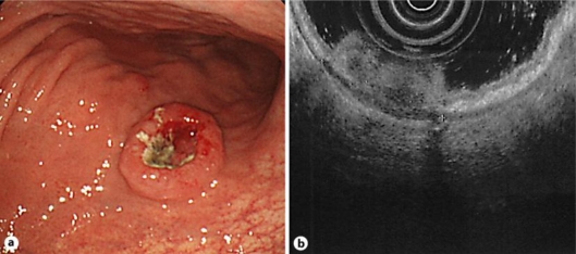
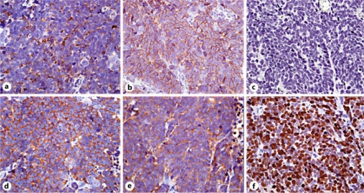
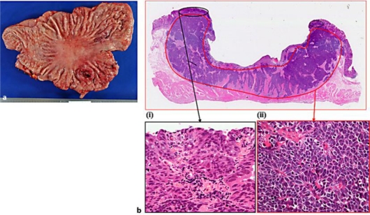
In the present study, we observed that the adenocarcinoma component in the mucosa was continuous with neuroendocrine carcinoma (NEC) in the deeper layers; this suggests the normal course of NEC carcinogenesis at the histological level. A 72-year-old man was admitted to our hospital with a chief complaint of tarry stools. Endoscopic examination of the upper gastrointestinal tract revealed a 2-cm tumor, with a deep central depression, surrounded by a smooth elevated area, in the middle of the stomach body. A biopsy showed that the tumor was a moderately differentiated gastric adenocarcinoma. The patient underwent total gastrectomy and standard lymph node dissection. The resected tumor was a 3.5 × 2.5 cm type 2 lesion. It comprised two elements at the histological level: (i) a moderately differentiated adenocarcinoma in the superficial portion of the mucous membrane layer, and (ii) NEC-like cells with dark, round nuclei and scant cytoplasm, presenting a solid and trabecular pattern, in the submucosal and muscularis propria layers. Immunohistochemical findings showed that the NEC-like cells were diffusely positive for chromogranin A, synaptophysin, neural cell adhesion molecule, and neuron-specific enolase, but were negative for carcinoembryonic antigen. The Ki-67 labeling index was 95%. The final pathological diagnosis was gastric NEC with an adenocarcinoma component and a high cellular proliferative potential.
在本研究中,我们观察到黏膜中的腺癌成分与深层的神经内分泌癌(NEC)连续;这在组织学水平提示了NEC的正常致癌过程。一名72岁男性因黑便为主诉入院。上消化道内镜检查发现胃体中部有一个2厘米的肿瘤,中央有一个深凹陷,周围是光滑的隆起区域。活检显示肿瘤为中度分化的胃腺癌。患者接受了全胃切除术和标准淋巴结清扫术。切除的肿瘤为3.5×2.5厘米的2型病变。在组织学水平上它由两个成分组成:(i)黏膜层浅表部分的中度分化腺癌,以及(ii)黏膜下层和固有肌层中具有深色圆形核和少量细胞质的NEC样细胞,呈实性和小梁状模式。免疫组化结果显示,NEC样细胞嗜铬粒蛋白A、突触素、神经细胞黏附分子和神经元特异性烯醇化酶弥漫性阳性,但癌胚抗原阴性。Ki-67标记指数为95%。最终病理诊断为伴有腺癌成分和高细胞增殖潜能的胃NEC。