Centre for Sports Medicine and Human Performance, Brunel University, Uxbridge, UK.
J Physiol. 2012 Jun 1;590(11):2767-82. doi: 10.1113/jphysiol.2012.228890. Epub 2012 Apr 2.
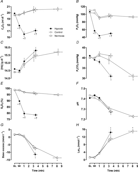
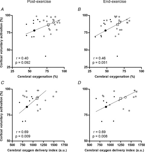
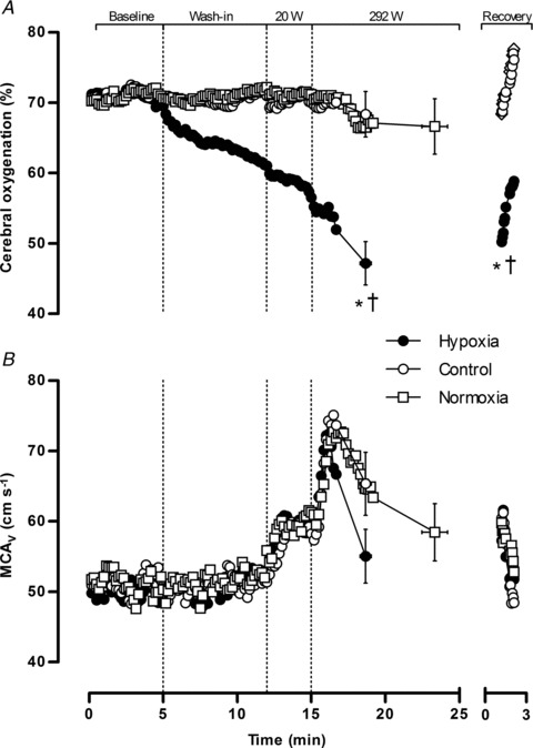
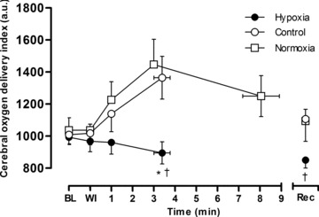
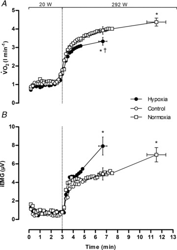
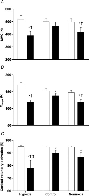
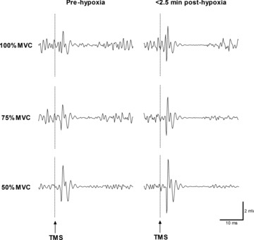
Inadequate cerebral O₂ availability has been proposed to be an important contributing factor to the development of central fatigue during strenuous exercise. Here we tested the hypothesis that supraspinal processes of fatigue would be increased after locomotor exercise in acute hypoxia compared to normoxia, and that such change would be related to reductions in cerebral O₂ delivery and tissue oxygenation. Nine endurance-trained cyclists completed three constant-load cycling exercise trials at ∼80% of maximal work rate: (1) to the limit of tolerance in acute hypoxia; (2) for the same duration but in normoxia (control); and (3) to the limit of tolerance in normoxia. Throughout each trial, prefrontal cortex tissue oxygenation and middle cerebral artery blood velocity (MCAV) were assessed using near-infrared spectroscopy and trans-cranial Doppler sonography, respectively. Cerebral O₂ delivery was calculated as the product of arterial O₂ content and MCAV. Before and immediately after each trial, twitch responses to supramaximal femoral nerve stimulation and transcranial magnetic stimulation were obtained to assess neuromuscular and cortical function, respectively. Exercise time was reduced by 54%in hypoxia compared to normoxia (3.6 ± 1.3 vs. 8.1 ± 2.9 min; P<0.001). Cerebral O₂ delivery,cerebral oxygenation and maximum O₂ uptake were reduced whereas muscle electromyographic activity was increased in hypoxia compared to control (P <0.05).Maximum voluntary force and potentiated quadriceps twitch force were decreased below baseline after exercise in each trial;the decreases were greater in hypoxia compared to control (P<0.001), but were not different in the exhaustive trials (P>0.05). Cortical voluntary activation was also decreased after exercise in all trials, but the decline in hypoxia (Δ18%) was greater than in the normoxic trials (Δ5-9%)(P <0.05). The reductions in cortical voluntary activation were paralleled by reductions in cerebral O₂ delivery. The results suggest that curtailment of exercise performance in acute severe hypoxia is due, in part, to failure of drive from the motor cortex, possibly as a consequence of diminished O₂ availability in the brain.
大脑供氧不足被认为是剧烈运动中中枢疲劳发展的一个重要因素。在这里,我们测试了一个假设,即在急性缺氧下进行运动后,会增加脊髓疲劳过程,并且这种变化与大脑氧输送和组织氧合减少有关。9 名耐力训练的自行车运动员在急性缺氧和常氧条件下完成了三种恒定负荷的自行车运动试验,约为最大工作率的 80%:(1)在急性缺氧下达到耐受极限;(2)在常氧下持续相同时间(对照);(3)在常氧下达到耐受极限。在每次试验中,通过近红外光谱法评估前额叶皮层组织氧合,通过经颅多普勒超声评估大脑中动脉血流速度(MCAV)。大脑氧输送被计算为动脉氧含量和 MCAV 的乘积。在每次试验前后,分别通过股神经最大刺激和经颅磁刺激获得肌电图和皮质功能的反应。与常氧相比,缺氧下的运动时间减少了 54%(3.6 ± 1.3 与 8.1 ± 2.9 分钟;P<0.001)。与对照相比,缺氧下的大脑氧输送、大脑氧合和最大摄氧量降低,而肌肉肌电图活动增加(P<0.05)。在每次试验后,最大自愿力和增强的股四头肌抽搐力均低于基线下降;与对照相比,缺氧下的下降更大(P<0.001),但在耗尽的试验中没有差异(P>0.05)。在所有试验后,皮质自愿激活也降低,但在缺氧下(下降 18%)大于常氧试验(下降 5-9%)(P<0.05)。皮质自愿激活的下降与大脑氧输送的减少平行。结果表明,在急性严重缺氧下运动表现的缩短部分归因于运动皮层驱动力的丧失,可能是由于大脑供氧不足所致。