Department of Molecular & Cellular Physiology, Louisiana State University Health Sciences Center, Shreveport, LA 71130, USA.
Semin Cell Dev Biol. 2012 Sep;23(7):729-37. doi: 10.1016/j.semcdb.2012.03.014. Epub 2012 Mar 30.
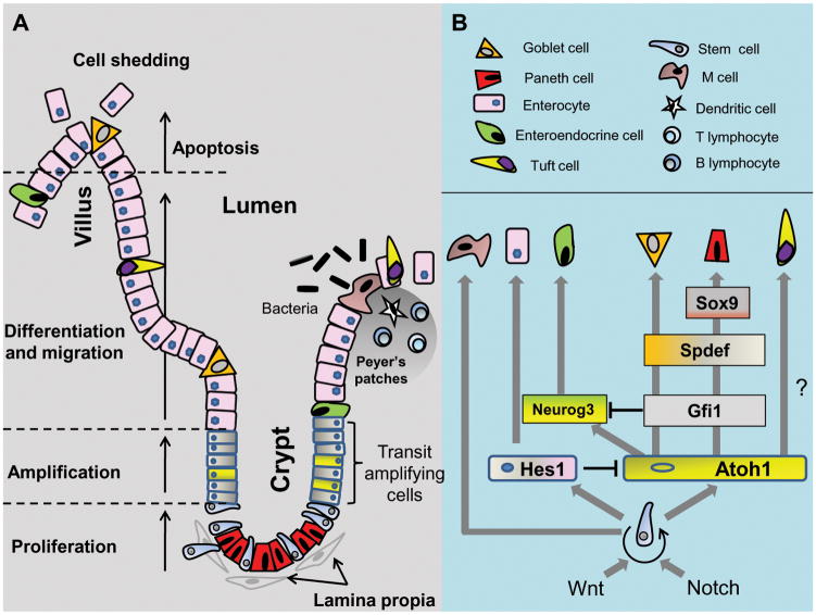
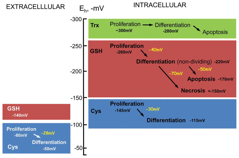
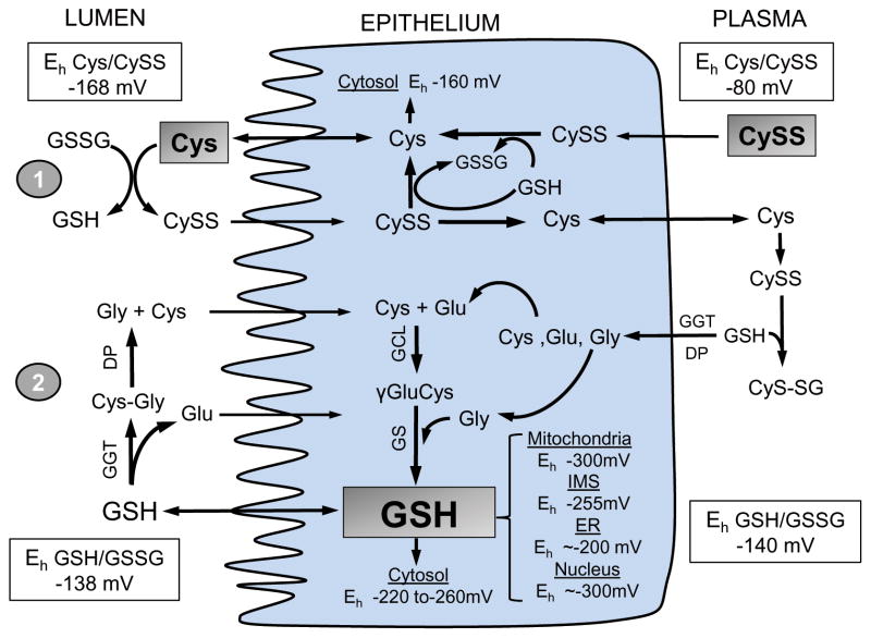
The intestinal epithelium sits at the interface between an organism and its luminal environment, and as such is prone to oxidative damage induced by luminal oxidants. Mucosal integrity is maintained by the luminal redox status of the glutathione/glutathione disulfide (GSH/GSSG) and cysteine/cystine (Cys/CySS) couples which also support luminal nutrient absorption, mucus fluidity, and a diverse microbiota. The epithelial layer is uniquely organized for rapid self-renewal that is achieved by the well-regulated processes of crypt stem cell proliferation and crypt-to-villus cell differentiation. The GSH/GSSG and Cys/CySS redox couples, known to modulate intestinal cell transition through proliferation, differentiation or apoptosis, could govern the regenerative potential of the mucosa. These two couples, together with that of the thioredoxin/thioredoxin disulfide (Trx/TrxSS) couple are the major intracellular redox systems, and it is proposed that they each function as distinctive redox control nodes or circuitry in the control of metabolic processes and networks of enzymatic reactions. Specificity of redox signaling is accomplished in part by subcellular compartmentation of the individual redox systems within the mitochondria, nucleus, endoplasmic reticulum, and cytosol wherein each defined redox environment is suited to the specific metabolic function within that compartment. Mucosal oxidative stress would result from the disruption of these unique redox control nodes, and the subsequent alteration in redox signaling can contribute to the development of degenerative pathologies of the intestine, such as inflammation and cancer.
肠上皮细胞位于生物体与其腔环境的交界处,因此容易受到腔内氧化剂诱导的氧化损伤。黏膜完整性由谷胱甘肽/谷胱甘肽二硫化物(GSH/GSSG)和半胱氨酸/胱氨酸(Cys/CySS)偶联物的腔内氧化还原状态维持,这些偶联物还支持腔内营养吸收、黏液流动性和多样化的微生物群。上皮层通过隐窝干细胞增殖和隐窝-绒毛细胞分化的精细调控过程进行快速自我更新,组织独特。已知 GSH/GSSG 和 Cys/CySS 氧化还原偶联物可通过增殖、分化或凋亡调节肠道细胞的转变,它们可能控制着黏膜的再生潜力。这两个偶联物,以及硫氧还蛋白/硫氧还蛋白二硫化物(Trx/TrxSS)偶联物,是主要的细胞内氧化还原系统,据推测,它们在控制代谢过程和酶反应网络方面各自作为独特的氧化还原控制节点或电路发挥作用。氧化还原信号的特异性部分是通过将各个氧化还原系统在细胞器(如线粒体、细胞核、内质网和细胞质)内进行亚细胞分隔来实现的,其中每个特定的氧化还原环境都适合该细胞器内的特定代谢功能。黏膜氧化应激会导致这些独特的氧化还原控制节点的破坏,随后的氧化还原信号改变可能导致肠道退行性病变的发展,如炎症和癌症。