Department of Radiology, 660 S. Euclid Ave, Campus Box 8131, Washington University School of Medicine, St Louis, MO 63110, USA.
Neuroimage. 2012 May 1;60(4):2182-90. doi: 10.1016/j.neuroimage.2012.02.044. Epub 2012 Feb 24.
Traditional T2 weighted MR imaging results are non-specific for the extent of underlying white matter structural abnormalities present in late life depression (LLD). Diffusion tensor imaging provides a unique opportunity to investigate the extent and nature of structural injury, but has been limited by examining only a subset of regions of interest (ROI) and by confounds common to the study of an elderly population, including comorbid vascular pathology. Furthermore, comprehensive correlation of diffusion tensor imaging (DTI) measurements, including axial and radial diffusivity measurements, has not been demonstrated in the late life depression population.
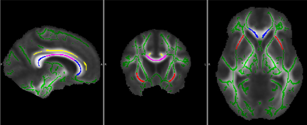
51 depressed and 16 non-depressed, age- and cerebrovascular risk factor-matched elderly subjects underwent traditional anatomic T1 and T2 weight imaging, as well as DTI. The DTI data were skeletonized using tract based spatial statistics (TBSS), and both regional and global analyses were performed.
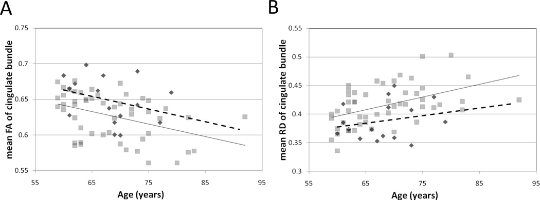
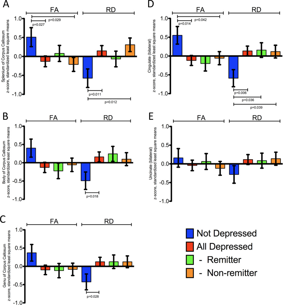
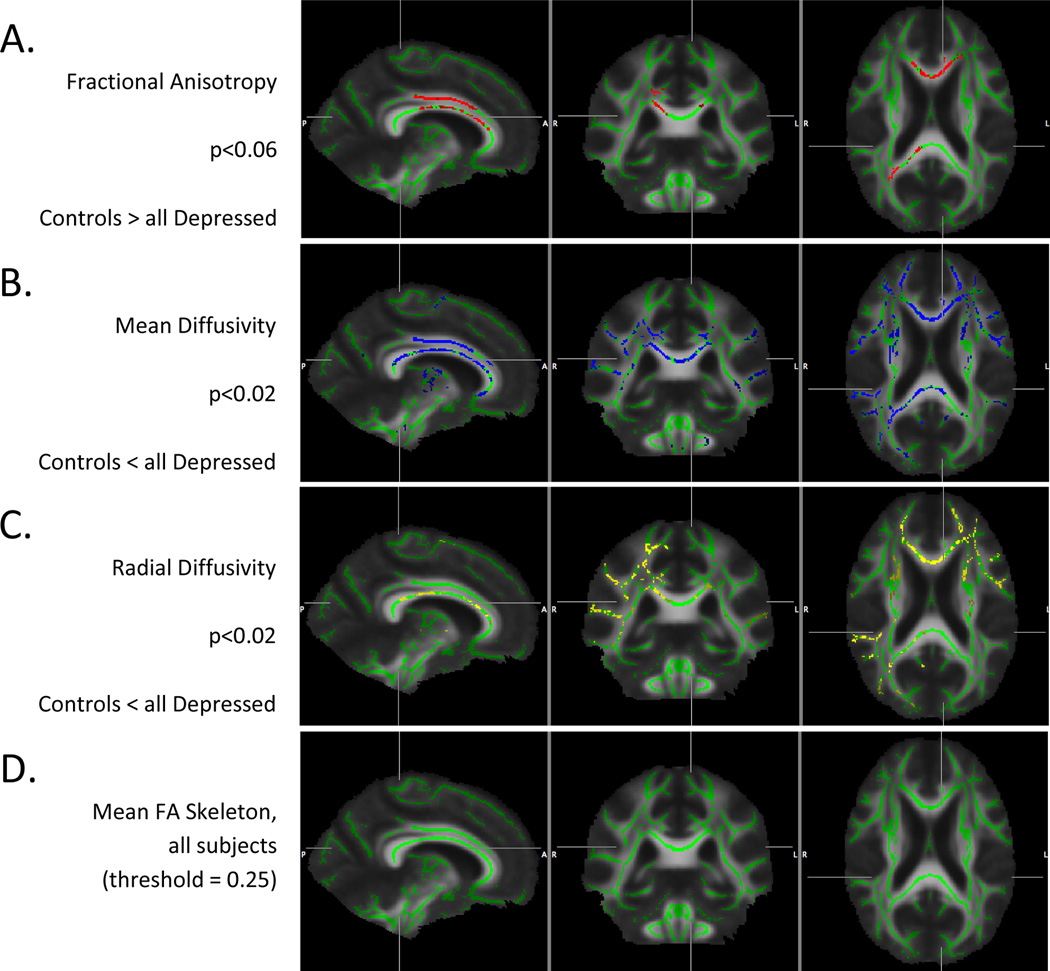
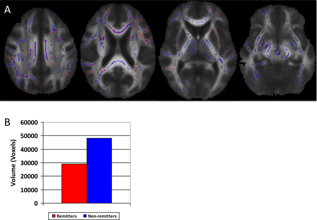
Widespread structural abnormalities within white matter were detected in the LLD group, accounting for age, gender and education and matched for cerebrovascular risk factors and global T2 white matter hyperintensities (T2WMH). Regional differences were most prominent in uncinate and cingulate white matter and were generally characterized by an increase in radial diffusivity. Age-related changes particularly in the cingulate bundle were more advanced in individuals with LLD relative to controls. Regression analysis demonstrated significant correlations of regional fractional anisotropy and radial diffusivity with five different neuropsychological factor scores. TBSS analysis demonstrated a greater extent of white matter abnormalities in LLD not responsive to treatment, as compared to controls.
White matter integrity is compromised in late life depression, largely manifested by increased radial diffusivity in specific regions, suggesting underlying myelin injury. A possible mechanism for underlying myelin injury is chronic white matter ischemia related to intrinsic cerebrovascular disease. In some regions such as the cingulate bundle, the white matter injury related to late life depression appears to be independent of and compounded by age-related changes. The correlations with neuropsychological testing indicate the essential effects of white matter injury on functional status. Lastly, response to treatment may depend on the extent of white matter injury, suggesting a need for intact functional networks.
传统的 T2 加权磁共振成像结果对于老年人抑郁症(LLD)中潜在的白质结构异常的程度是非特异性的。弥散张量成像提供了一个独特的机会来研究结构损伤的程度和性质,但由于只检查了一部分感兴趣区域(ROI),以及由于研究老年人群体中常见的混杂因素,包括合并的血管病理学,受到了限制。此外,弥散张量成像(DTI)测量的综合相关性,包括轴向和径向弥散度测量,在老年抑郁症人群中尚未得到证明。
51 名抑郁和 16 名非抑郁、年龄和脑血管危险因素匹配的老年受试者接受了传统的解剖 T1 和 T2 加权成像以及 DTI。DTI 数据使用基于束的空间统计学(TBSS)进行骨架化,进行了区域和全局分析。
在 LLD 组中检测到广泛的白质内结构异常,这些异常与年龄、性别和教育有关,并与脑血管危险因素和全局 T2 白质高信号(T2WMH)相匹配。区域差异在钩回和扣带回白质中最为明显,通常表现为径向弥散度增加。与对照组相比,LLD 患者的年龄相关变化,特别是在扣带束中,更为先进。回归分析显示,局部各向异性分数和径向弥散度与五个不同的神经心理学因素评分显著相关。TBSS 分析显示,与对照组相比,治疗无反应的 LLD 患者的白质异常程度更大。
老年人抑郁症的白质完整性受损,主要表现为特定区域的径向弥散度增加,提示存在脱髓鞘损伤。脱髓鞘损伤的潜在机制可能是与内在脑血管疾病相关的慢性白质缺血。在一些区域,如扣带束,与老年人抑郁症相关的白质损伤似乎独立于年龄相关的变化,并且与之叠加。与神经心理学测试的相关性表明白质损伤对功能状态的重要影响。最后,治疗反应可能取决于白质损伤的程度,这表明需要完整的功能网络。