• 文献检索
  • 文档翻译
  • 深度研究
  • 学术资讯
  • Suppr Zotero 插件Zotero 插件
  • 邀请有礼
  • 套餐&价格
  • 历史记录
应用&插件
Suppr Zotero 插件Zotero 插件浏览器插件Mac 客户端Windows 客户端微信小程序
定价
高级版会员购买积分包购买API积分包
服务
文献检索文档翻译深度研究API 文档MCP 服务
关于我们
关于 Suppr公司介绍联系我们用户协议隐私条款
关注我们

Suppr 超能文献

核心技术专利:CN118964589B侵权必究
粤ICP备2023148730 号-1Suppr @ 2026

文献检索

告别复杂PubMed语法,用中文像聊天一样搜索,搜遍4000万医学文献。AI智能推荐,让科研检索更轻松。

立即免费搜索

文件翻译

保留排版,准确专业,支持PDF/Word/PPT等文件格式,支持 12+语言互译。

免费翻译文档

深度研究

AI帮你快速写综述,25分钟生成高质量综述,智能提取关键信息,辅助科研写作。

立即免费体验

贝伐珠单抗治疗复发性高级别脑肿瘤患者的早期灌注变化:定量评估的初步结果。

Early perfusion changes in patients with recurrent high-grade brain tumor treated with Bevacizumab: preliminary results by a quantitative evaluation.

机构信息

Radiology and Diagnostic Imaging Department, Regina Elena Cancer Institute, Via Elio Chianesi 53, 00144, Rome, Italy.

出版信息

J Exp Clin Cancer Res. 2012 Apr 11;31(1):33. doi: 10.1186/1756-9966-31-33.

DOI:10.1186/1756-9966-31-33
PMID:22494770
原文链接:https://pmc.ncbi.nlm.nih.gov/articles/PMC3583244/
Abstract

BACKGROUND

To determine whether early monitoring of the effects of bevacizumab in patients with recurrent high-grade gliomas, by a Perfusion Computed Tomography (PCT), may be a predictor of the response to treatment assessed through conventional MRI follow-up.

METHODS

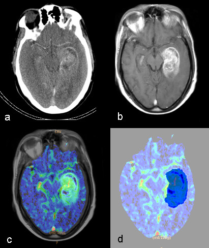
Sixteen patients were enrolled in the present study. For each patient, two PCT examinations, before and after the first dose of bevacizumab, were acquired. Areas of abnormal Cerebral Blood Volume (CBV) were manually defined on the CBV maps, using co-registered T1- weighted images, acquired before treatment, as a guide to the tumor location. Different perfusion metrics were derived from the histogram analysis of the normalized CBV (nCBV) maps; both hyper and hypo-perfused sub-volumes were quantified in the lesion, including tumor necrosis. A two-tailed Wilcoxon test was used to establish the significance of changes in the different perfusion metrics, observed at baseline and during treatment. The relationships between changes in perfusion and morphological MRI modifications at first follow-up were investigated.

RESULTS

Significant reductions in mean and median nCBV were detected throughout the entire patient population, after only a single dose of bevacizumab. The nCBV histogram modifications indicated the normalization effect of bevacizumab on the tumor abnormal vasculature. An improvement in hypoxia after a single dose of bevacizumab was predictive of a greater reduction in T1-weighted contrast-enhanced volumes at first follow-up.

CONCLUSIONS

These preliminary results show that a quantification of changes in necrotic intra-tumoral regions could be proposed as a potential imaging biomarker of tumor response to anti-VEGF therapies.

摘要

背景

通过灌注计算机断层扫描(PCT)来早期监测复发性高级别胶质瘤患者贝伐单抗的疗效,这种方法是否可以作为通过常规 MRI 随访评估治疗反应的预测因子。

方法

本研究纳入了 16 名患者。每位患者在接受贝伐单抗首剂量之前和之后分别进行了两次 PCT 检查。使用经治疗前 T1 加权图像配准的 CBV 图,手动定义异常脑血容量(CBV)区域,以作为肿瘤位置的指导。从归一化 CBV(nCBV)图的直方图分析中得出了不同的灌注指标;在病变中量化了高灌注和低灌注的亚体积,包括肿瘤坏死。采用双尾 Wilcoxon 检验来确定在基线和治疗期间观察到的不同灌注指标的变化的显著性。研究了灌注变化与首次随访时的形态学 MRI 改变之间的关系。

结果

在整个患者群体中,仅接受单次贝伐单抗治疗后,平均和中位数 nCBV 均显著降低。nCBV 直方图的改变表明贝伐单抗对肿瘤异常血管的正常化作用。单次贝伐单抗治疗后缺氧的改善可预测首次随访时 T1 加权对比增强体积的更大减少。

结论

这些初步结果表明,对坏死性肿瘤内区域变化的定量可以作为肿瘤对抗血管生成治疗反应的潜在影像学生物标志物。

https://cdn.ncbi.nlm.nih.gov/pmc/blobs/79ea/3583244/13e9943aed80/1756-9966-31-33-4.jpg
https://cdn.ncbi.nlm.nih.gov/pmc/blobs/79ea/3583244/71d6a285dd32/1756-9966-31-33-1.jpg
https://cdn.ncbi.nlm.nih.gov/pmc/blobs/79ea/3583244/2bd791da9b83/1756-9966-31-33-2.jpg
https://cdn.ncbi.nlm.nih.gov/pmc/blobs/79ea/3583244/007ae3df5780/1756-9966-31-33-3.jpg
https://cdn.ncbi.nlm.nih.gov/pmc/blobs/79ea/3583244/13e9943aed80/1756-9966-31-33-4.jpg

相似文献

1
Early perfusion changes in patients with recurrent high-grade brain tumor treated with Bevacizumab: preliminary results by a quantitative evaluation.贝伐珠单抗治疗复发性高级别脑肿瘤患者的早期灌注变化:定量评估的初步结果。
J Exp Clin Cancer Res. 2012 Apr 11;31(1):33. doi: 10.1186/1756-9966-31-33.
2
Early biomarkers from dynamic contrast-enhanced magnetic resonance imaging to predict the response to antiangiogenic therapy in high-grade gliomas.基于动态对比增强磁共振成像的早期生物标志物预测高级别胶质瘤抗血管生成治疗反应
Neuroradiology. 2015 Dec;57(12):1269-80. doi: 10.1007/s00234-015-1582-9. Epub 2015 Sep 12.
3
Recurrent glioblastoma treated with bevacizumab: contrast-enhanced T1-weighted subtraction maps improve tumor delineation and aid prediction of survival in a multicenter clinical trial.贝伐珠单抗治疗复发性胶质母细胞瘤:多中心临床试验中,对比增强 T1 加权减影图可改善肿瘤勾画,有助于预测生存。
Radiology. 2014 Apr;271(1):200-10. doi: 10.1148/radiol.13131305. Epub 2013 Nov 27.
4
Leakage decrease detected by dynamic susceptibility-weighted contrast-enhanced perfusion MRI predicts survival in recurrent glioblastoma treated with bevacizumab.动态对比增强磁共振成像检测到的渗漏减少可预测接受贝伐单抗治疗的复发性胶质母细胞瘤患者的生存期。
Clin Transl Oncol. 2017 Jan;19(1):51-57. doi: 10.1007/s12094-016-1502-4. Epub 2016 Mar 30.
5
Whole-tumor histogram analysis of the cerebral blood volume map: tumor volume defined by 11C-methionine positron emission tomography image improves the diagnostic accuracy of cerebral glioma grading.脑血容量图的全肿瘤直方图分析:由11C-蛋氨酸正电子发射断层扫描图像定义的肿瘤体积可提高脑胶质瘤分级的诊断准确性。
Jpn J Radiol. 2017 Oct;35(10):613-621. doi: 10.1007/s11604-017-0675-2. Epub 2017 Sep 6.
6
Dynamic-susceptibility contrast agent MRI measures of relative cerebral blood volume predict response to bevacizumab in recurrent high-grade glioma.动态对比剂磁共振成像测量相对脑血容量可预测复发性高级别胶质瘤对贝伐珠单抗的反应。
Neuro Oncol. 2014 Jun;16(6):880-8. doi: 10.1093/neuonc/not216. Epub 2014 Jan 15.
7
Vascular change measured with independent component analysis of dynamic susceptibility contrast MRI predicts bevacizumab response in high-grade glioma.采用动态磁敏感对比 MRI 的独立成分分析测量血管变化可预测高级别胶质瘤对贝伐单抗的反应。
Neuro Oncol. 2013 Apr;15(4):442-50. doi: 10.1093/neuonc/nos323. Epub 2013 Feb 3.
8
Identification of Early Response to Anti-Angiogenic Therapy in Recurrent Glioblastoma: Amide Proton Transfer-weighted and Perfusion-weighted MRI compared with Diffusion-weighted MRI.复发性脑胶质瘤抗血管生成治疗早期反应的识别:酰胺质子转移加权和灌注加权 MRI 与弥散加权 MRI 的比较。
Radiology. 2020 May;295(2):397-406. doi: 10.1148/radiol.2020191376. Epub 2020 Mar 10.
9
Quantitative T2 mapping of recurrent glioblastoma under bevacizumab improves monitoring for non-enhancing tumor progression and predicts overall survival.贝伐单抗治疗复发性胶质母细胞瘤的定量 T2 映射可改善对非增强肿瘤进展的监测,并预测总生存期。
Neuro Oncol. 2013 Oct;15(10):1395-404. doi: 10.1093/neuonc/not105. Epub 2013 Aug 7.
10
Treatment with bevacizumab plus carboplatin for recurrent malignant glioma.贝伐珠单抗联合卡铂治疗复发性恶性脑胶质瘤。
Neurosurgery. 2010 Jul;67(1):87-93. doi: 10.1227/01.NEU.0000370918.51053.BC.

引用本文的文献

1
Baseline T1 hyperintense and diffusion-restricted lesions are not linked to prolonged survival in bevacizumab-treated glioblastoma patients of the GLARIUS trial.基线 T1 高信号和弥散受限病变与 GLARIUS 试验贝伐珠单抗治疗的胶质母细胞瘤患者的生存延长无关。
J Neurooncol. 2019 Sep;144(3):501-509. doi: 10.1007/s11060-019-03246-4. Epub 2019 Jul 19.
2
Predicting Glioblastoma Response to Bevacizumab Through MRI Biomarkers of the Tumor Microenvironment.通过肿瘤微环境的 MRI 生物标志物预测胶质母细胞瘤对贝伐珠单抗的反应。
Mol Imaging Biol. 2019 Aug;21(4):747-757. doi: 10.1007/s11307-018-1289-5.
3
Vascular Hysteresis Loops and Vascular Architecture Mapping in Patients with Glioblastoma treated with Antiangiogenic Therapy.

本文引用的文献

1
Bevacizumab impairs oxidative energy metabolism and shows antitumoral effects in recurrent glioblastomas: a 31P/1H MRSI and quantitative magnetic resonance imaging study.贝伐单抗抑制复发性胶质母细胞瘤的氧化能量代谢并显示抗肿瘤作用:一项 31P/1H MRSI 和定量磁共振成像研究。
Neuro Oncol. 2011 Dec;13(12):1349-63. doi: 10.1093/neuonc/nor132. Epub 2011 Sep 2.
2
Computed tomography perfusion imaging for therapeutic assessment: has it come of age as a biomarker in oncology?计算机断层灌注成像在治疗评估中的应用:它是否已成为肿瘤学中的一种生物标志物?
Invest Radiol. 2012 Jan;47(1):2-4. doi: 10.1097/RLI.0b013e318229ff3e.
3
Reducing intratumour acute hypoxia through bevacizumab treatment, referring to the response of quiescent tumour cells and metastatic potential.
抗血管生成治疗的胶质母细胞瘤患者的血管迟滞环和血管结构图谱
Sci Rep. 2017 Aug 17;7(1):8508. doi: 10.1038/s41598-017-09048-w.
4
Glioma FMISO PET/MR Imaging Concurrent with Antiangiogenic Therapy: Molecular Imaging as a Clinical Tool in the Burgeoning Era of Personalized Medicine.胶质瘤氟代脱氧葡萄糖正电子发射断层显像/磁共振成像与抗血管生成治疗同步进行:在蓬勃发展的个性化医疗时代,分子成像作为一种临床工具
Biomedicines. 2016 Oct 31;4(4):24. doi: 10.3390/biomedicines4040024.
5
Monitoring the Response of Hyperbilirubinemia in the Mouse Brain by In Vivo Bioluminescence Imaging.通过活体生物发光成像监测小鼠脑中高胆红素血症的反应。
Int J Mol Sci. 2016 Dec 28;18(1):50. doi: 10.3390/ijms18010050.
6
Misleading early blood volume changes obtained using ferumoxytol-based magnetic resonance imaging perfusion in high grade glial neoplasms treated with bevacizumab.使用基于铁羧麦芽糖的磁共振成像灌注技术在接受贝伐单抗治疗的高级别胶质肿瘤中获得的早期血容量变化存在误导性。
Fluids Barriers CNS. 2016 Dec 20;13(1):23. doi: 10.1186/s12987-016-0047-9.
7
Quantitative Perfusion and Permeability Biomarkers in Brain Cancer from Tomographic CT and MR Images.基于断层CT和MR图像的脑癌定量灌注与渗透性生物标志物
Biomark Cancer. 2016 Jul 3;8(Suppl 2):47-59. doi: 10.4137/BIC.S31801. eCollection 2016.
8
Additive antiangiogenesis effect of ginsenoside Rg3 with low-dose metronomic temozolomide on rat glioma cells both in vivo and in vitro.人参皂苷Rg3与低剂量节拍性替莫唑胺对大鼠胶质瘤细胞的体内外联合抗血管生成作用。
J Exp Clin Cancer Res. 2016 Feb 13;35:32. doi: 10.1186/s13046-015-0274-y.
9
Early biomarkers from dynamic contrast-enhanced magnetic resonance imaging to predict the response to antiangiogenic therapy in high-grade gliomas.基于动态对比增强磁共振成像的早期生物标志物预测高级别胶质瘤抗血管生成治疗反应
Neuroradiology. 2015 Dec;57(12):1269-80. doi: 10.1007/s00234-015-1582-9. Epub 2015 Sep 12.
10
Advanced MR Imaging in Neuro-oncology.神经肿瘤学中的先进磁共振成像
Clin Neuroradiol. 2015 Oct;25 Suppl 2:143-9. doi: 10.1007/s00062-015-0439-2. Epub 2015 Jul 29.
通过贝伐单抗治疗减少肿瘤内急性缺氧,这是指静止肿瘤细胞的反应和转移潜力。
Br J Radiol. 2011 Dec;84(1008):1131-8. doi: 10.1259/bjr/38457938. Epub 2011 May 17.
4
Perfusion CT findings in patients with metastatic carcinoid tumors undergoing bevacizumab and interferon therapy.转移性类癌肿瘤患者接受贝伐珠单抗和干扰素治疗的灌注 CT 表现。
AJR Am J Roentgenol. 2011 Mar;196(3):569-76. doi: 10.2214/AJR.10.4455.
5
Quantitative volumetric analysis of conventional MRI response in recurrent glioblastoma treated with bevacizumab.贝伐珠单抗治疗复发性胶质母细胞瘤的常规 MRI 反应的定量容积分析。
Neuro Oncol. 2011 Apr;13(4):401-9. doi: 10.1093/neuonc/noq206. Epub 2011 Feb 15.
6
Anti-VEGF treatment reduces blood supply and increases tumor cell invasion in glioblastoma.抗血管内皮生长因子治疗减少胶质母细胞瘤的血液供应并增加肿瘤细胞浸润。
Proc Natl Acad Sci U S A. 2011 Mar 1;108(9):3749-54. doi: 10.1073/pnas.1014480108. Epub 2011 Feb 14.
7
Antiangiogenic therapy for primary liver cancer: correlation of changes in dynamic contrast-enhanced magnetic resonance imaging with tissue hypoxia markers and clinical response.原发性肝癌的抗血管生成治疗:动态对比增强磁共振成像与组织缺氧标志物的变化与临床反应的相关性。
Ann Surg Oncol. 2011 Aug;18(8):2192-9. doi: 10.1245/s10434-011-1570-1. Epub 2011 Feb 1.
8
Early evaluation of tumoral response to antiangiogenic therapy by arterial spin labeling perfusion magnetic resonance imaging and susceptibility weighted imaging in a patient with recurrent glioblastoma receiving bevacizumab.在一名接受贝伐单抗治疗的复发性胶质母细胞瘤患者中,通过动脉自旋标记灌注磁共振成像和磁敏感加权成像对肿瘤对抗血管生成治疗的反应进行早期评估。
J Clin Oncol. 2011 Apr 10;29(11):e308-11. doi: 10.1200/JCO.2010.32.6082. Epub 2011 Jan 24.
9
ADC histograms predict response to anti-angiogenic therapy in patients with recurrent high-grade glioma.ADC 直方图预测复发性高级别胶质瘤患者对抗血管生成治疗的反应。
Neuroradiology. 2011 Apr;53(4):291-302. doi: 10.1007/s00234-010-0808-0. Epub 2010 Dec 2.
10
Scale to predict survival after surgery for recurrent glioblastoma multiforme.预测复发性多形性胶质母细胞瘤手术后生存的评分。
J Clin Oncol. 2010 Aug 20;28(24):3838-43. doi: 10.1200/JCO.2010.30.0582. Epub 2010 Jul 19.