Manni Isabella, Di Rocco Giuliana, Fusco Salvatore, Leone Lucia, Barbati Saviana Antonella, Carapella Carmine Maria, Grassi Claudio, Piaggio Giulia, Toietta Gabriele
Department of Research, Advanced Diagnostic, and Technological Innovation, Regina Elena National Cancer Institute, 00144 Rome, Italy.
Institute of Human Physiology, Medical School, Università Cattolica del Sacro Cuore, 00168 Rome, Italy.
Int J Mol Sci. 2016 Dec 28;18(1):50. doi: 10.3390/ijms18010050.
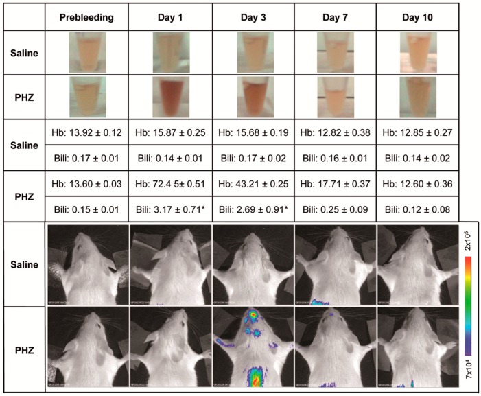
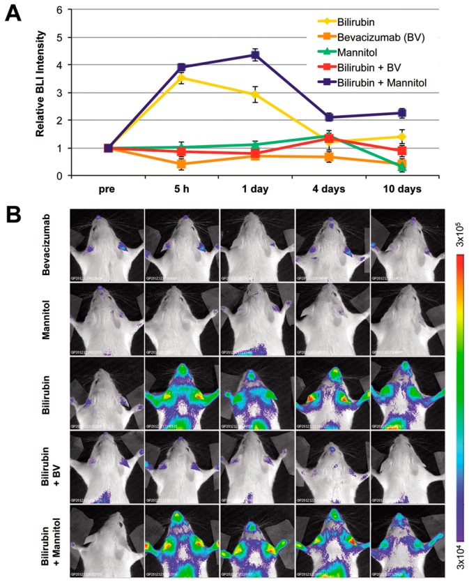
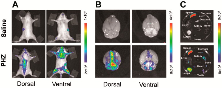
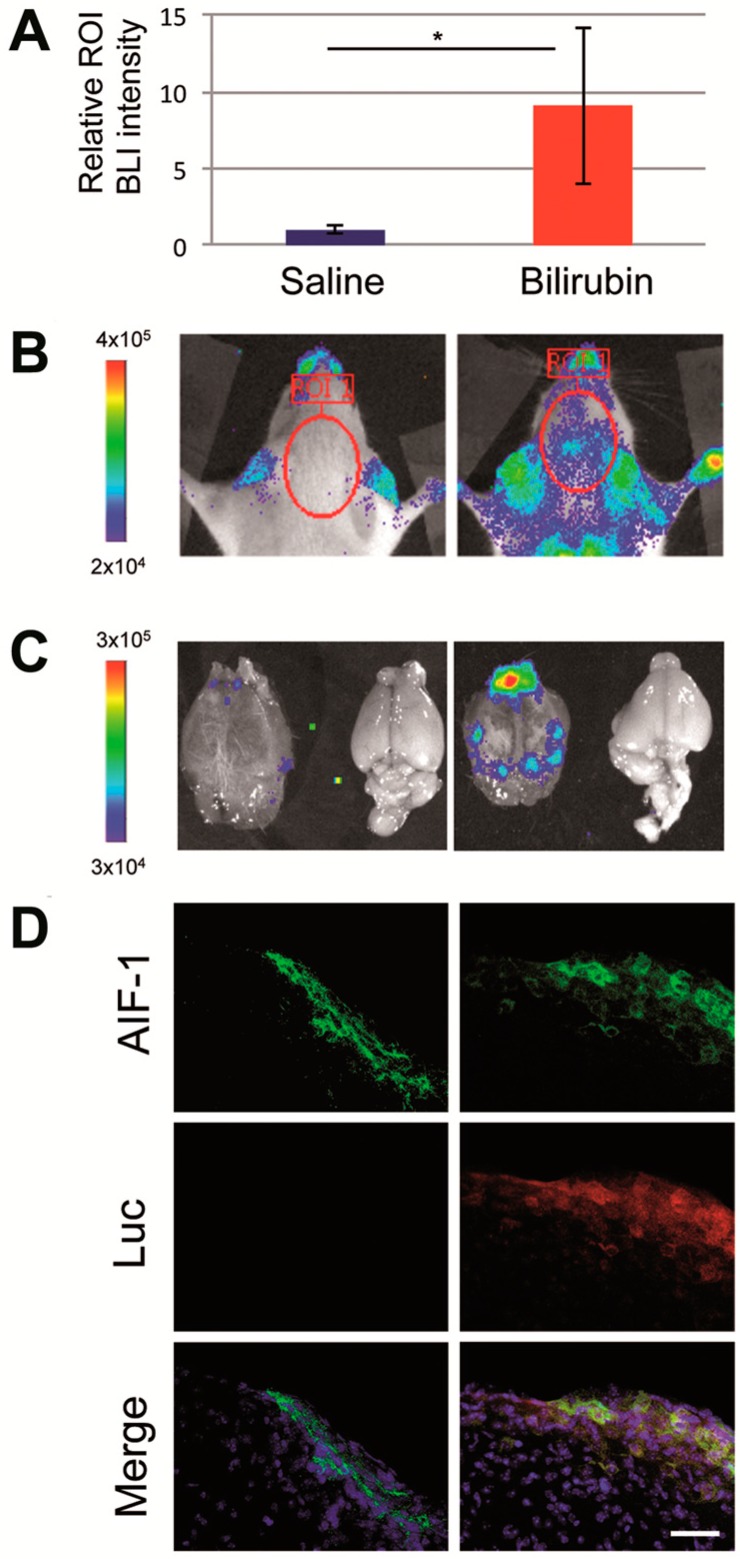
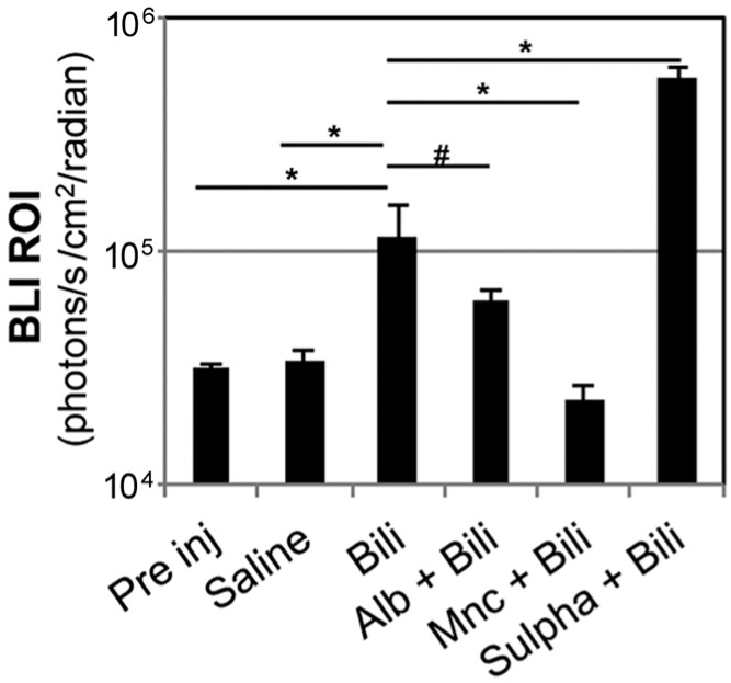
Increased levels of unconjugated bilirubin are neurotoxic, but the mechanism leading to neurological damage has not been completely elucidated. Innovative strategies of investigation are needed to more precisely define this pathological process. By longitudinal in vivo bioluminescence imaging, we noninvasively visualized the brain response to hyperbilirubinemia in the MITO-Luc mouse, in which light emission is restricted to the regions of active cell proliferation. We assessed that acute hyperbilirubinemia promotes bioluminescence in the brain region, indicating an increment in the cell proliferation rate. Immunohistochemical detection in brain sections of cells positive for both luciferase and the microglial marker allograft inflammatory factor 1 suggests proliferation of microglial cells. In addition, we demonstrated that brain induction of bioluminescence was altered by pharmacological displacement of bilirubin from its albumin binding sites and by modulation of the blood-brain barrier permeability, all pivotal factors in the development of bilirubin-induced neurologic dysfunction. We also determined that treatment with minocycline, an antibiotic with anti-inflammatory and neuroprotective properties, or administration of bevacizumab, an anti-vascular endothelial growth factor antibody, blunts bilirubin-induced bioluminescence. Overall the study supports the use of the MITO-Luc mouse as a valuable tool for the rapid response monitoring of drugs aiming at preventing acute bilirubin-induced neurological dysfunction.
未结合胆红素水平升高具有神经毒性,但导致神经损伤的机制尚未完全阐明。需要创新的研究策略来更精确地界定这一病理过程。通过纵向活体生物发光成像,我们在MITO-Luc小鼠中对大脑对高胆红素血症的反应进行了无创可视化,在该小鼠中,光发射仅限于活跃细胞增殖区域。我们评估发现,急性高胆红素血症会促进大脑区域的生物发光,表明细胞增殖率增加。对脑切片中同时对荧光素酶和小胶质细胞标志物同种异体移植炎症因子1呈阳性的细胞进行免疫组织化学检测,提示小胶质细胞增殖。此外,我们证明,胆红素从其白蛋白结合位点的药理学置换以及血脑屏障通透性的调节会改变大脑生物发光的诱导,这些都是胆红素诱导神经功能障碍发展中的关键因素。我们还确定,使用具有抗炎和神经保护特性的抗生素米诺环素治疗,或给予抗血管内皮生长因子抗体贝伐单抗,可减弱胆红素诱导的生物发光。总体而言,该研究支持将MITO-Luc小鼠作为一种有价值的工具,用于快速监测旨在预防急性胆红素诱导神经功能障碍的药物的反应。