Yu Qi, Li Yafeng, Waqar Ahmed Bilal, Wang Yanli, Huang Bingqiao, Chen Yulong, Zhao Sihai, Yang Peigang, Fan Jianglin, Liu Enqi
Research Institute of Atherosclerotic Disease, Xi'an Jiaotong University School of Medicine, Xi'an 710061, China.
J Biomed Biotechnol. 2012;2012:506159. doi: 10.1155/2012/506159. Epub 2012 Mar 14.
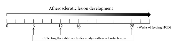
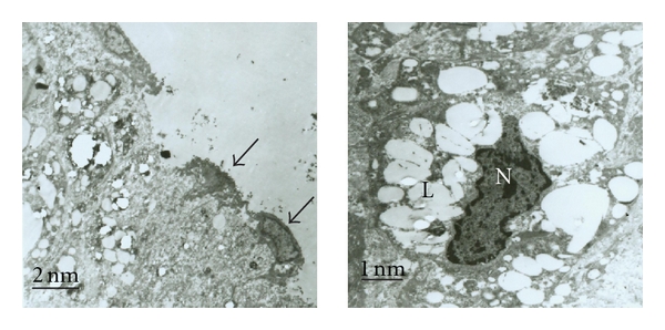
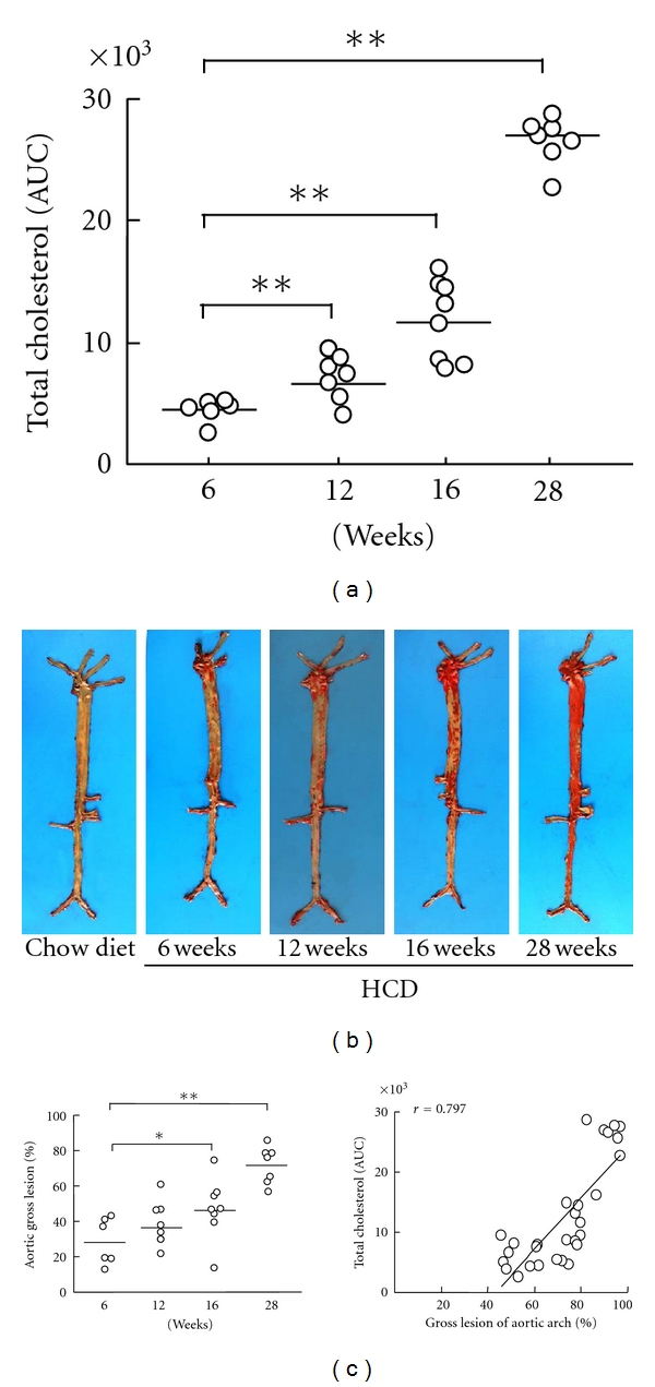
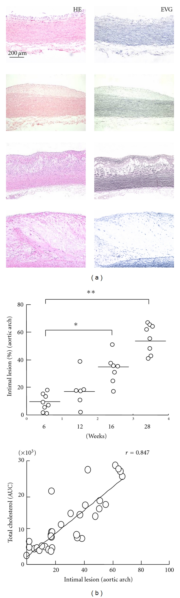
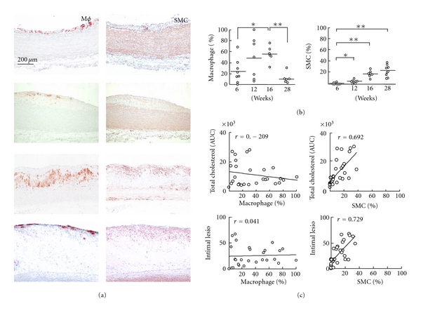
The diet-induced atherosclerotic rabbit is an ideal model for atherosclerosis study, but temporal changes in atherosclerotic development in hypercholesterolemic rabbits are poorly understood. Japanese white rabbits were fed a high-cholesterol diet to induce sustained hypercholesterolemia, and each group of 10-12 animals was then sacrificed at 6, 12, 16, or 28 weeks. The rabbit aortas were harvested, and the sizes of the gross and intima atherosclerotic lesions were quantified. The cellular component of macrophages (Mφs) and smooth muscle cells (SMCs) in aortic intimal lesions was also quantified by immunohistochemical staining, and the correlation between plasma cholesterol levels and the progress of atherosclerotic lesions was studied. The ultrastructure of the atherosclerotic lesions was observed by transmission electron microscopy (TEM). Widely variable atherosclerotic plaques were found from 6 weeks to 28 weeks, and the lesional progress was closely correlated with cholesterol exposure. Interestingly, a relatively reduced accumulation of Mφ, an increased numbers of SMCs, and a damaged endothelial layer were presented in advanced lesions. Moreover, SMCs were closely correlated with cholesterol exposure and lesional progress for the whole period. Cholesterol exposure directly determines atherosclerotic progress in a rabbit model, and the changes in the cellular component of advanced lesions may affect plaque stability in an atherosclerotic rabbit model.
饮食诱导的动脉粥样硬化兔是动脉粥样硬化研究的理想模型,但高胆固醇血症兔动脉粥样硬化发展的时间变化尚不清楚。给日本白兔喂食高胆固醇饮食以诱导持续性高胆固醇血症,然后每组10 - 12只动物在6、12、16或28周时处死。采集兔主动脉,对大体和内膜动脉粥样硬化病变的大小进行量化。还通过免疫组织化学染色对主动脉内膜病变中巨噬细胞(Mφs)和平滑肌细胞(SMCs)的细胞成分进行量化,并研究血浆胆固醇水平与动脉粥样硬化病变进展之间的相关性。通过透射电子显微镜(TEM)观察动脉粥样硬化病变的超微结构。在6周龄至28周龄的兔中发现了广泛变化的动脉粥样硬化斑块,病变进展与胆固醇暴露密切相关。有趣的是,在晚期病变中出现了相对减少的Mφ积累、增加的SMC数量和受损的内皮细胞层。此外,在整个时期内,SMC与胆固醇暴露和病变进展密切相关。胆固醇暴露直接决定了兔模型中的动脉粥样硬化进展,晚期病变细胞成分的变化可能会影响动脉粥样硬化兔模型中斑块的稳定性。