Department of Radiology, University of Arizona, Tucson, AZ, USA.
Mol Imaging. 2012 Jun;11(3):187-96. doi: 10.2310/7290.2011.00039.
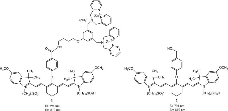
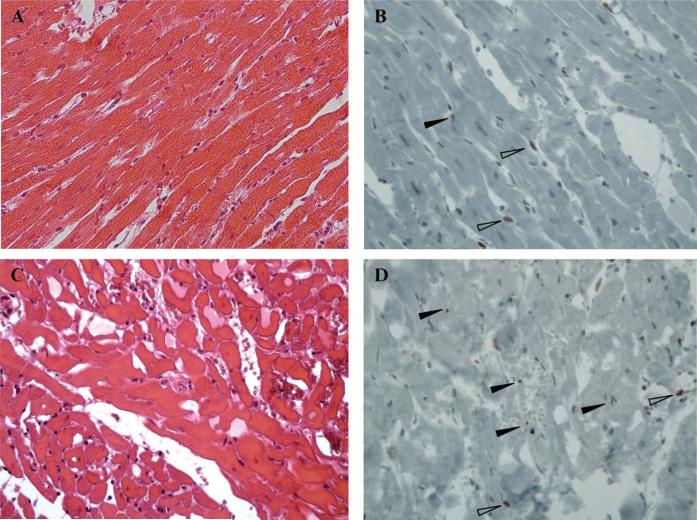
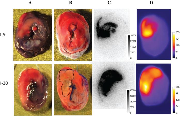
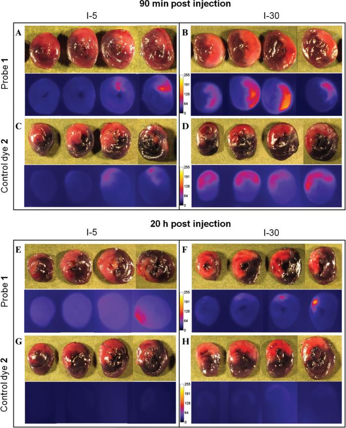
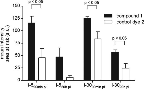
A fluorescent zinc 2,2'-dipicolylamine coordination complex PSVue®794 (probe 1) is known to selectively bind to phosphatidylserine exposed on the surface of apoptotic and necrotic cells. In this study, we investigated the cell death targeting properties of probe 1 in myocardial ischemia-reperfusion injury. A rat heart model of ischemia-reperfusion was used. Probe 1, control dye, or 99mTc glucarate was intravenously injected in rats subjected to 30-minute and 5-minute myocardial ischemia followed by 2-hour reperfusion. At 90 minutes or 20 hours postinjection, myocardial uptake was evaluated ex vivo by fluorescence imaging and autoradiography. Hematoxylin-eosin and cleaved caspase-3 staining was performed on myocardial sections to demonstrate the presence of ischemia-reperfusion injury and apoptosis. Selective accumulation of probe 1 could be detected in the area at risk up to 20 hours postinjection. Similar topography and extent of uptake of probe 1 and 99mTc glucarate were observed at 90 minutes postinjection. Histologic analysis demonstrated the presence of necrosis, but only a few apoptotic cells could be detected. Probe 1 selectively accumulates in myocardial ischemia-reperfusion injury and is a promising cell death imaging tool.
一种荧光锌 2,2'-二吡啶甲酰胺配合物 PSVue®794(探针 1)已知可选择性结合在凋亡和坏死细胞表面暴露的磷脂酰丝氨酸上。在这项研究中,我们研究了探针 1 在心肌缺血再灌注损伤中的细胞死亡靶向特性。使用大鼠心肌缺血再灌注模型。在经历 30 分钟和 5 分钟心肌缺血,然后再灌注 2 小时的大鼠中静脉注射探针 1、对照染料或 99mTc 葡萄糖酸盐。在注射后 90 分钟或 20 小时,通过荧光成像和放射自显影术评估心肌的摄取。对心肌切片进行苏木精-伊红和裂解的 caspase-3 染色,以证明存在缺血再灌注损伤和细胞凋亡。在注射后 20 小时内,可在危险区域检测到探针 1 的选择性积聚。在注射后 90 分钟,观察到探针 1 和 99mTc 葡萄糖酸盐的摄取具有相似的形态和范围。组织学分析表明存在坏死,但只能检测到少数凋亡细胞。探针 1 选择性地积聚在心肌缺血再灌注损伤中,是一种很有前途的细胞死亡成像工具。