Division of Gastroenterology and Hepatology, The University of Alabama at Birmingham, Birmingham, Alabama, Lebanon.
Ann Thorac Med. 2012 Apr;7(2):84-91. doi: 10.4103/1817-1737.94527.
Mediastinal lymphadenopathy (ML) is a cause for concern, especially in patients with previous malignancy. We report our experience with the use of endoscopic ultrasound-guided fine needle aspiration (EUS-FNA) with immunocytochemical stains in patients being evaluated for ML.
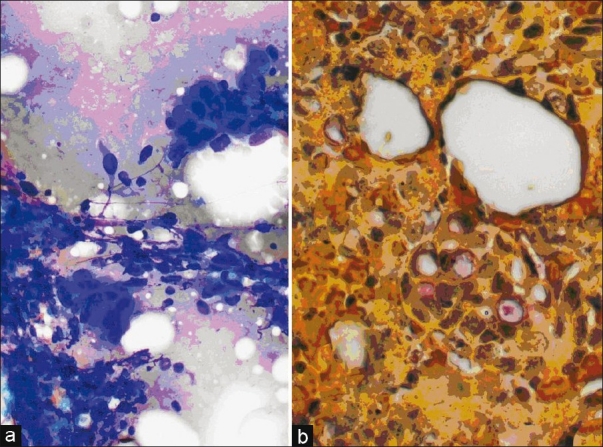
Retrospective analysis of patients with ML of unknown origin who underwent EUS-FNA. On-site evaluation was performed by experienced cytologist, and special immunocytochemical stains were requested as indicated.
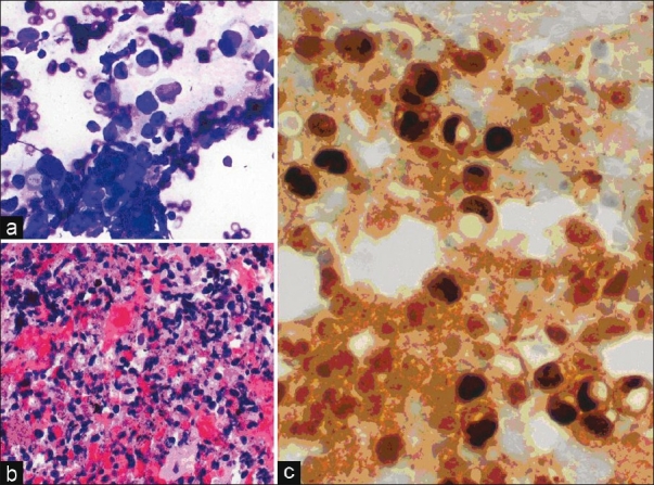
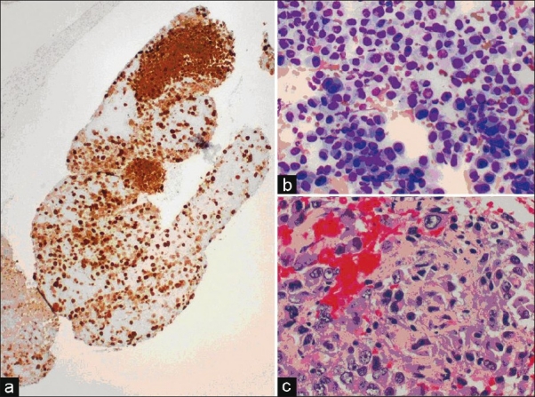
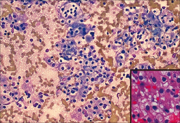
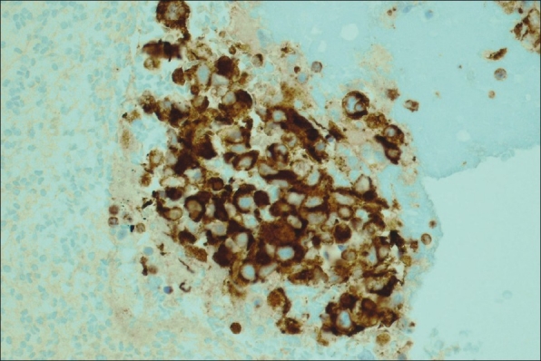
A total of 116 patients were included, and a total of 136 mediastinal LN were sampled. Prior malignancy was present in 45%. The most common site of examined lymph node (LN) were subcarinal (76%, 103 LN). The median long and short axis diameters were 28 mm and 13 mm, respectively. FNA was read on-site as malignant, 21 (16%); benign, 100 (76.9%); suspicious, six (4%); atypical, 3 (2%); and inadequate sample, six (4%). Sixty-four LN were deferred for additional studies; 22 for immunocytochemical and 26 for Gimesa (GMS) stain and 21 for flow cytometry. Final FNA read was malignant in 28 (21%), benign in 103 (76%), suspicious in three (2%), and atypical in two (1%). Metastatic malignancies disclosed included Hodgkin's and Non-Hodgkin's lymphoma, melanoma, hepatoma, breast, lung, colon, renal, endometrial, Fallopian tube, and unknown carcinoma. The sensitivity, specificity, and accuracy of the final FNA read to predict malignancy were 100%.
EUS-guided FNA with additional ancillary studies is useful in disclosing metastatic ML from a variety of neoplasms. Due to its safety and accuracy profile, it should be considered the test of choice in evaluating abnormal ML in appropriately selected patients.
纵隔淋巴结病(ML)是一个值得关注的问题,特别是在有既往恶性肿瘤病史的患者中。我们报告了在评估 ML 患者时使用内镜超声引导下细针抽吸(EUS-FNA)联合免疫细胞化学染色的经验。
回顾性分析因不明原因纵隔淋巴结病而行 EUS-FNA 的患者。由经验丰富的细胞学家进行现场评估,并根据需要请求特殊免疫细胞化学染色。
共纳入 116 例患者,共对 136 个纵隔淋巴结进行了采样。45%的患者有既往恶性肿瘤病史。最常检查的淋巴结部位是隆突下(76%,103 个淋巴结)。淋巴结的长轴和短轴直径中位数分别为 28mm 和 13mm。现场 FNA 判读结果为恶性 21 例(16%);良性 100 例(76.9%);可疑 6 例(4%);不典型 3 例(2%);标本不足 6 例(4%)。64 个淋巴结需要进一步研究;22 个进行免疫细胞化学染色,26 个进行 Gimesa(GMS)染色,21 个进行流式细胞术。最终 FNA 判读结果为恶性 28 例(21%),良性 103 例(76%),可疑 3 例(2%),不典型 2 例(1%)。揭示的转移性恶性肿瘤包括霍奇金和非霍奇金淋巴瘤、黑色素瘤、肝癌、乳腺癌、肺癌、结肠癌、肾癌、子宫内膜癌、输卵管癌和未知来源的癌。最终 FNA 判读预测恶性肿瘤的灵敏度、特异性和准确性均为 100%。
EUS 引导下 FNA 联合辅助检查有助于揭示来自多种肿瘤的转移性 ML。由于其安全性和准确性,在适当选择的患者中,它应被视为评估异常纵隔淋巴结的首选检查。