Aschendorff Antje
HNO-Klinik und Implant Centrum Freiburg, Universität Freiburg, Germany.
GMS Curr Top Otorhinolaryngol Head Neck Surg. 2011;10:Doc07. doi: 10.3205/cto000080. Epub 2012 Apr 26.
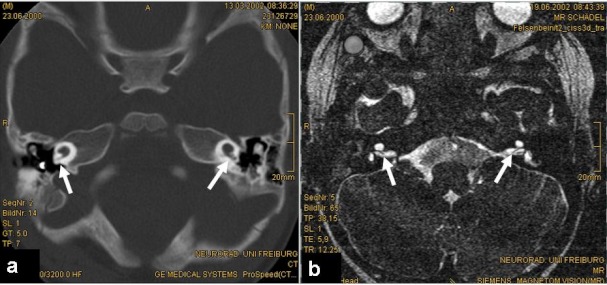
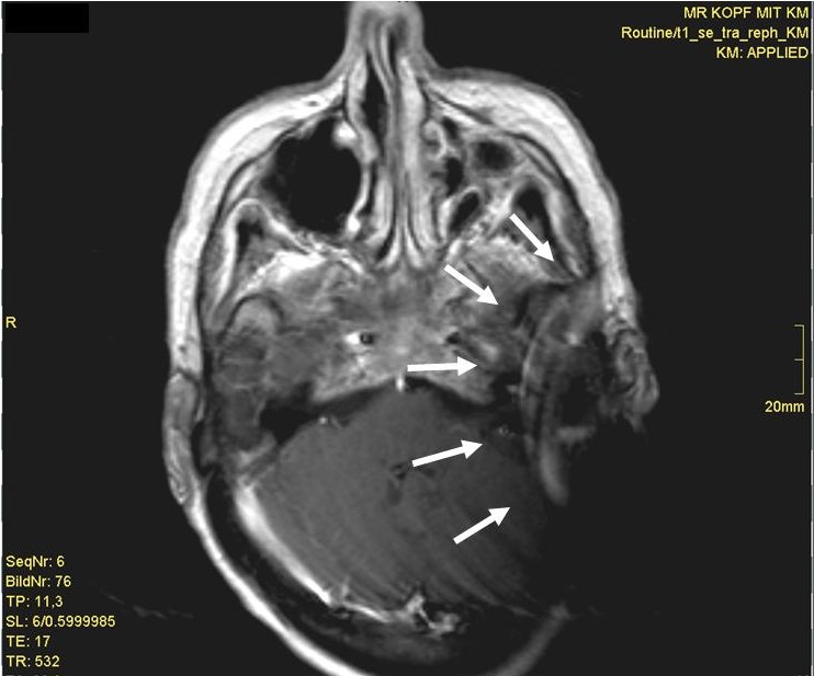
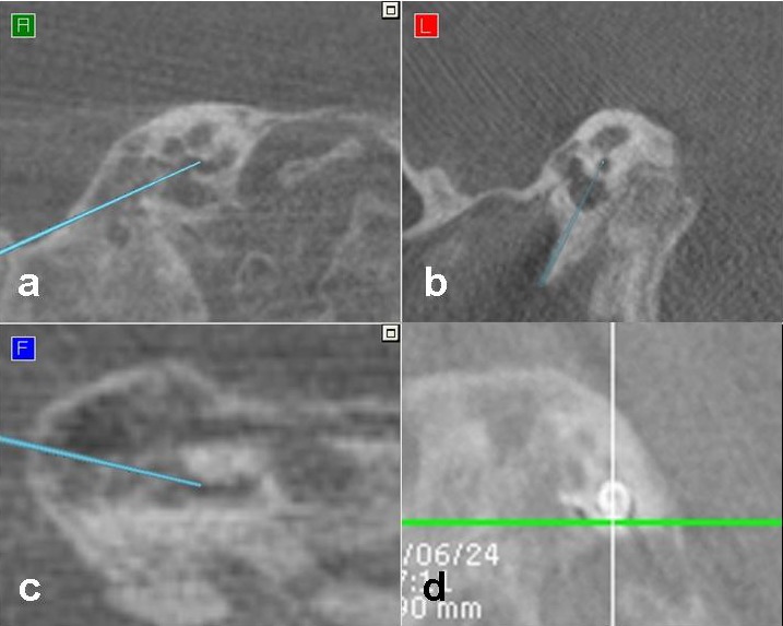
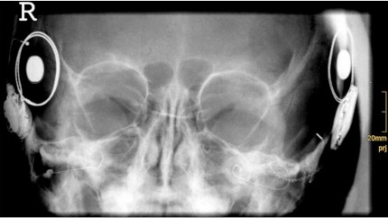
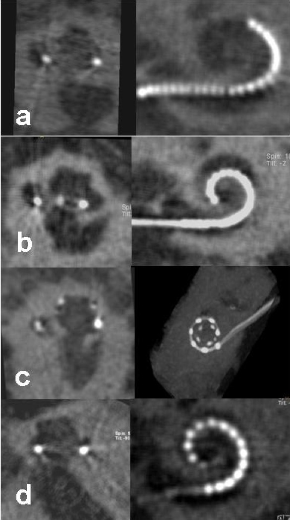
Imaging procedures are a mainstream tool in the daily ENT workflow. Cochlear Implant patients are representing a special population with specific demands for imaging. There are different imaging techniques available for pre-operative evaluation, surgery and postoperative controls with different indications and consequences. High-resolution computed tomography and magnetic resonance imaging are mainly used in the evaluation process. New procedures, as digital volume tomography, are increasingly used intra- and postoperatively. Especially the intracochlear positioning in malformations of the inner ear, eventually added with radiological assisted navigation, can be considered a standard of modern cochlear implant surgery. In addition, digital volume tomography may serve as a quality control tool focusing on the evaluation of the intracochlear electrode position. The range of applications, indications and current results are illustrated.
成像程序是耳鼻喉科日常工作流程中的主流工具。人工耳蜗植入患者是对成像有特殊需求的特殊人群。有多种不同的成像技术可用于术前评估、手术及术后检查,其适应证和结果各不相同。高分辨率计算机断层扫描和磁共振成像主要用于评估过程。新的程序,如数字容积断层扫描,在手术中和术后越来越多地被使用。特别是在内耳畸形中人工耳蜗的耳蜗内定位,最终辅以放射学辅助导航,可被视为现代人工耳蜗植入手术的标准。此外,数字容积断层扫描可作为一种质量控制工具,专注于评估人工耳蜗电极在耳蜗内的位置。文中阐述了其应用范围、适应证及当前的结果。