Research Service (151), Veterans Affairs Medical Center, 113 Holland Ave, Albany, NY, 12208, USA.
Cerebellum. 2012 Dec;11(4):845-60. doi: 10.1007/s12311-012-0383-5.
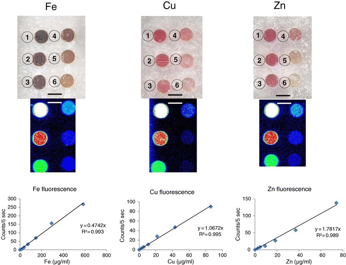
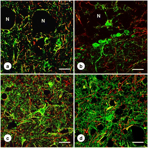
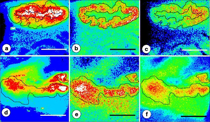
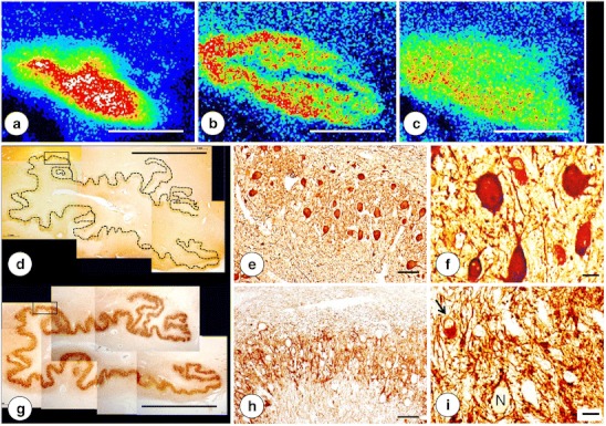
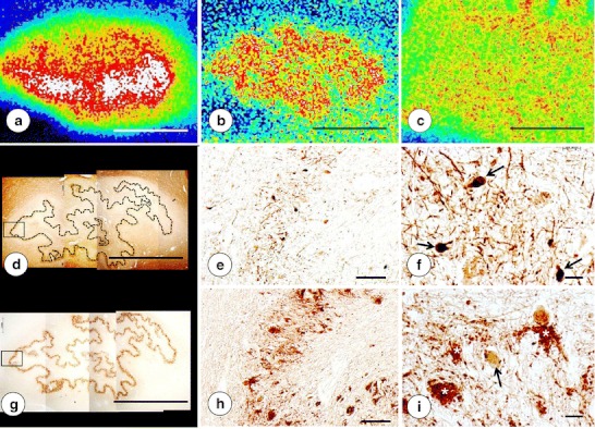
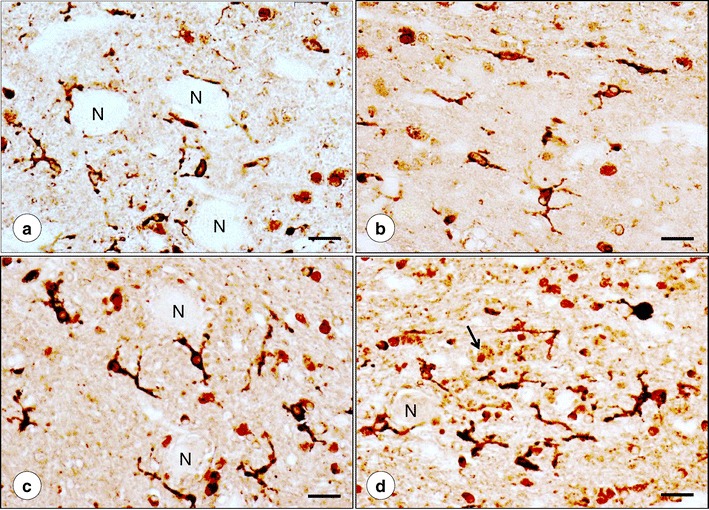
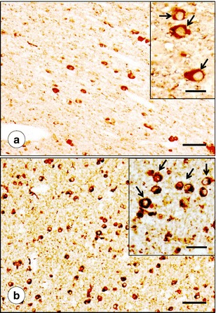
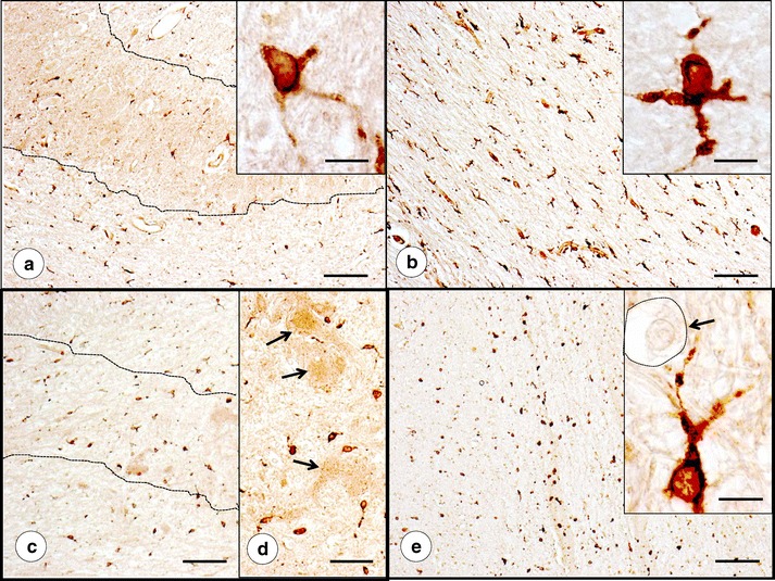
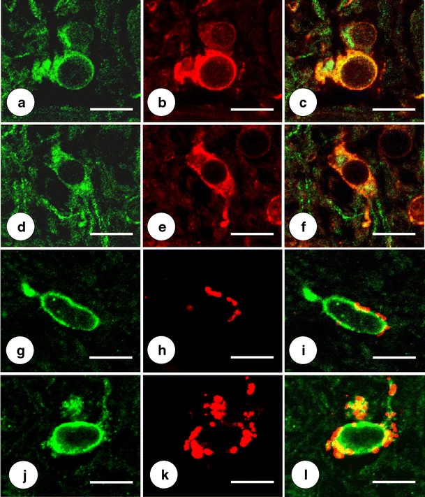
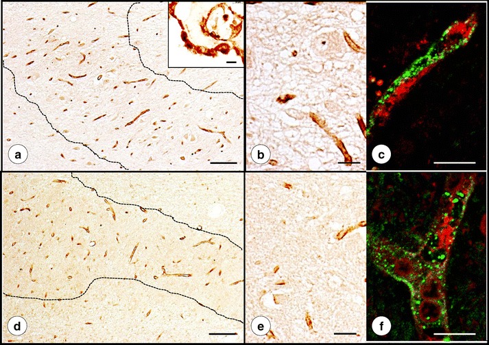
Friedreich's ataxia (FRDA) causes selective atrophy of the large neurons of the dentate nucleus (DN). High iron (Fe) concentration and failure to clear the metal from the affected brain tissue are potential risk factors in the progression of the lesion. The DN also contains relatively high amounts of copper (Cu) and zinc (Zn), but the importance of these metals in FRDA has not been established. This report describes nondestructive quantitative X-ray fluorescence (XRF) and "mapping" of Fe, Cu, and Zn in polyethylene glycol-dimethylsulfoxide (PEG/DMSO)-embedded DN of 10 FRDA patients and 13 controls. Fe fluorescence arose predominantly from the hilar white matter, whereas Cu and Zn were present at peak levels in DN gray matter. Despite collapse of the DN in FRDA, the location of the peak Fe signal did not change. In contrast, the Cu and Zn regions broadened and overlapped extensively with the Fe-rich region. Maximal metal concentrations did not differ from normal (in micrograms per milliliter of solid PEG/DMSO as means ± S.D.): Fe normal, 364 ± 117, FRDA, 344 ± 159; Cu normal, 33 ± 13, FRDA, 33 ± 18; and Zn normal, 32 ± 16, FRDA, 33 ± 19. Tissues were recovered from PEG/DMSO and transferred into paraffin for matching with immunohistochemistry of neuron-specific enolase (NSE), glutamic acid decarboxylase (GAD), and ferritin. NSE and GAD reaction products confirmed neuronal atrophy and grumose degeneration that coincided with abnormally diffuse Cu and Zn zones. Ferritin immunohistochemistry matched Fe XRF maps, revealing the most abundant reaction product in oligodendroglia of the DN hilus. In FRDA, these cells were smaller and more numerous than normal. In the atrophic DN gray matter of FRDA, anti-ferritin labeled mostly hypertrophic microglia. Immunohistochemistry and immunofluorescence of the Cu-responsive proteins Cu,Zn-superoxide dismutase and Cu(++)-transporting ATPase α-peptide did not detect specific responses to Cu redistribution in FRDA. In contrast, metallothionein (MT)-positive processes were more abundant than normal and contributed to the gliosis of the DN. The isoforms of MT, MT-1/2, and brain-specific MT-3 displayed only limited co-localization with glial fibrillary acidic protein. The results suggest that MT can provide effective protection against endogenous Cu and Zn toxicity in FRDA, similar to the neuroprotective sequestration of Fe in holoferritin.
弗里德里希共济失调症 (FRDA) 导致齿状核 (DN) 的大神经元选择性萎缩。铁 (Fe) 浓度高且未能从受影响的脑组织中清除金属是病变进展的潜在危险因素。DN 还含有相对较高量的铜 (Cu) 和锌 (Zn),但这些金属在 FRDA 中的重要性尚未确定。本报告描述了用聚乙二醇-二甲基亚砜 (PEG/DMSO) 包埋的 10 名 FRDA 患者和 13 名对照的 DN 进行无损定量 X 射线荧光 (XRF) 和“映射”的 Fe、Cu 和 Zn。Fe 荧光主要来自齿状核白质的神经细胞,而 Cu 和 Zn 则在 DN 灰质中达到峰值水平。尽管 FRDA 中 DN 塌陷,但最大 Fe 信号的位置没有改变。相比之下,Cu 和 Zn 区域广泛扩大并与富含 Fe 的区域重叠。最大金属浓度与正常情况无差异(以每毫升固态 PEG/DMSO 中的微克数表示平均值±SD):Fe 正常,364±117,FRDA,344±159;Cu 正常,33±13,FRDA,33±18;Zn 正常,32±16,FRDA,33±19。将组织从 PEG/DMSO 中回收并转移到石蜡中,与神经元特异性烯醇化酶 (NSE)、谷氨酸脱羧酶 (GAD) 和铁蛋白的免疫组化相匹配。NSE 和 GAD 反应产物证实了神经元萎缩和颗粒状变性,与异常弥散的 Cu 和 Zn 区域相吻合。铁蛋白免疫组化与 Fe XRF 图谱相匹配,在齿状核神经细胞体的神经胶质细胞中发现了最丰富的反应产物。在 FRDA 中,这些细胞比正常情况下更小、更多。在 FRDA 萎缩的 DN 灰质中,抗铁蛋白标记的主要是肥大的小胶质细胞。Cu,Zn-超氧化物歧化酶和 Cu(++)-转运 ATP 酶 α-肽的 Cu 反应蛋白的免疫组化和免疫荧光检测未发现 FRDA 中 Cu 再分布的特异性反应。相比之下,金属硫蛋白 (MT)-阳性过程比正常情况下更为丰富,并促成了 DN 的神经胶质增生。MT 的同工型 MT-1/2 和脑特异性 MT-3 仅与神经胶质纤维酸性蛋白有有限的共定位。结果表明,MT 可以为 FRDA 提供有效的内源性 Cu 和 Zn 毒性保护,类似于铁蛋白中铁的神经保护螯合。