Top Institute Food and Nutrition, Wageningen, The Netherlands.
PLoS One. 2012;7(4):e35008. doi: 10.1371/journal.pone.0035008. Epub 2012 Apr 19.
Intestinal barrier dysfunction and translocation of endotoxins are involved in the pathogenesis of alcoholic liver disease. Exposure to ethanol and its metabolite, acetaldehyde at relatively high concentrations have been shown to disrupt intestinal epithelial tight junctions in the conventional two dimensional cell culture models. The present study investigated quantitatively and qualitatively the effects of ethanol at concentrations detected in the blood after moderate ethanol consumption, of its metabolite acetaldehyde and of the combination of both compounds on intestinal barrier function in a three-dimensional cell culture model.
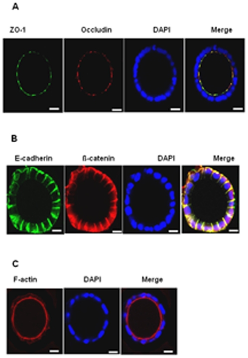
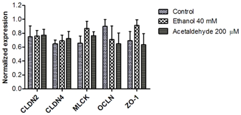
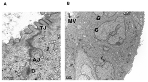
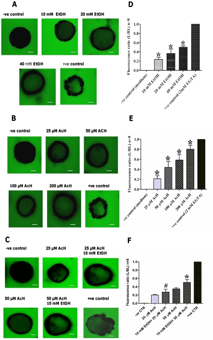
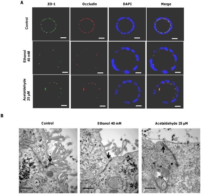
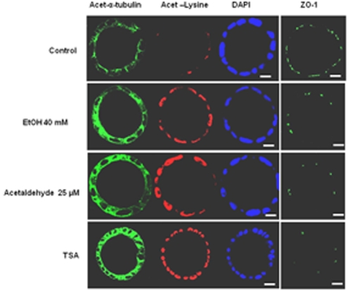
Caco-2 cells were grown in a basement membrane matrix (Matrigel™) to induce spheroid formation and were then exposed to the compounds at the basolateral side. Morphological differentiation of the spheroids was assessed by immunocytochemistry and transmission electron microscopy. The barrier function was assessed by the flux of FITC-labeled dextran from the basal side into the spheroids' luminal compartment using confocal microscopy. Caco-2 cells grown on Matrigel assembled into fully differentiated and polarized spheroids with a central lumen, closely resembling enterocytes in vivo and provide an excellent model to study epithelial barrier functionality. Exposure to ethanol (10-40 mM) or acetaldehyde (25-200 µM) for 3 h, dose-dependently and additively increased the paracellular permeability and induced redistribution of ZO-1 and occludin without affecting cell viability or tight junction-encoding gene expression. Furthermore, ethanol and acetaldehyde induced lysine residue and microtubules hyperacetylation.
These results indicate that ethanol at concentrations found in the blood after moderate drinking and acetaldehyde, alone and in combination, can increase the intestinal epithelial permeability. The data also point to the involvement of protein hyperacetylation in ethanol- and acetaldehyde-induced loss of tight junctions integrity.
肠道屏障功能障碍和内毒素易位参与了酒精性肝病的发病机制。研究表明,在传统的二维细胞培养模型中,乙醇及其代谢产物乙醛在相对较高的浓度下暴露会破坏肠道上皮细胞紧密连接。本研究在三维细胞培养模型中定量和定性研究了中等饮酒量后血液中检测到的乙醇、其代谢产物乙醛以及这两种化合物的组合对肠道屏障功能的影响。
Caco-2 细胞在基底膜基质(Matrigel™)中生长以诱导球体形成,然后在基底外侧暴露于化合物。通过免疫细胞化学和透射电子显微镜评估球体的形态分化。通过共聚焦显微镜评估 FITC 标记的葡聚糖从基底侧进入球体腔室的通量来评估屏障功能。在 Matrigel 上生长的 Caco-2 细胞组装成完全分化和极化的球体,具有中央腔室,与体内肠细胞非常相似,为研究上皮屏障功能提供了极好的模型。暴露于乙醇(10-40mM)或乙醛(25-200µM)3 小时,呈剂量依赖性和累加性地增加了细胞旁通透性,并导致 ZO-1 和闭合蛋白重新分布,而不影响细胞活力或紧密连接编码基因的表达。此外,乙醇和乙醛诱导赖氨酸残基和微管过度乙酰化。
这些结果表明,在中等饮酒后血液中发现的乙醇浓度以及单独和联合的乙醛可增加肠道上皮通透性。这些数据还表明,蛋白质过度乙酰化参与了乙醇和乙醛诱导的紧密连接完整性丧失。