Computer-Assisted Drug Design Lab, Research Programme on Biomedical Informatics (GRIB), PRBB, Dr Aiguader 88, Barcelona, 08003, Spain.
J Mol Model. 2012 Sep;18(9):4465-75. doi: 10.1007/s00894-012-1431-2. Epub 2012 May 29.
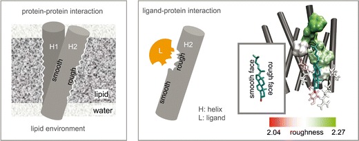
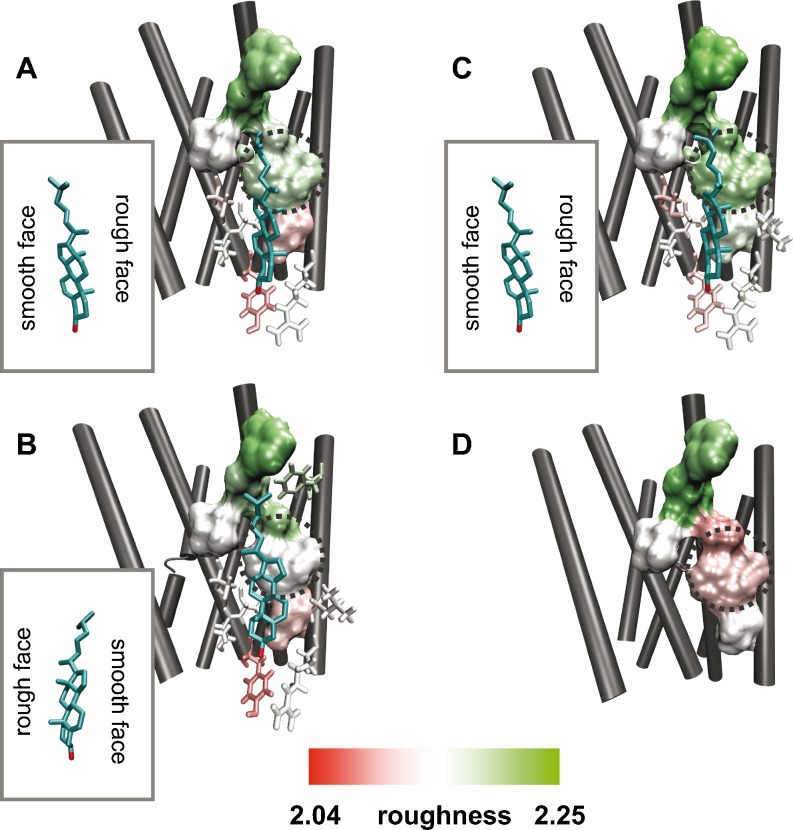
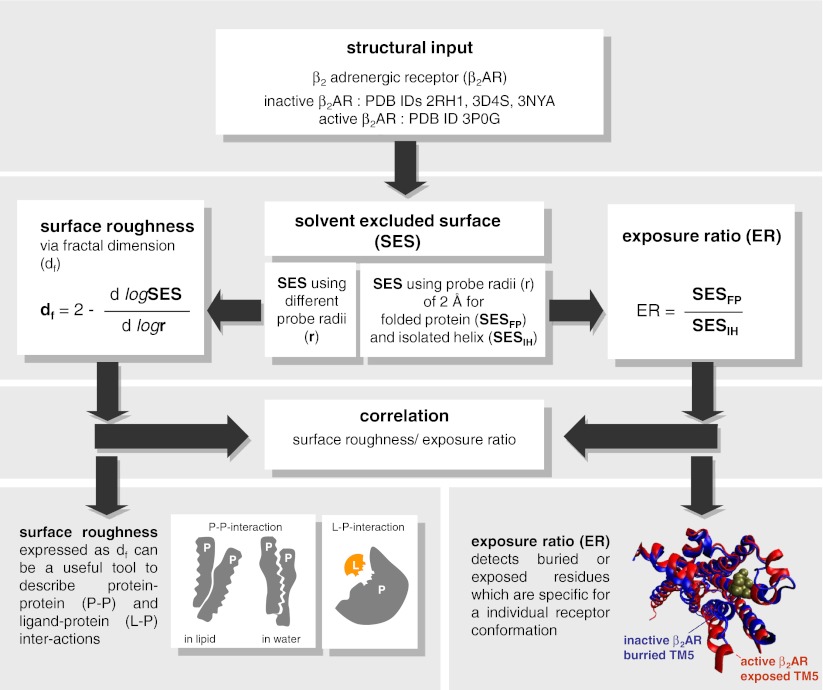
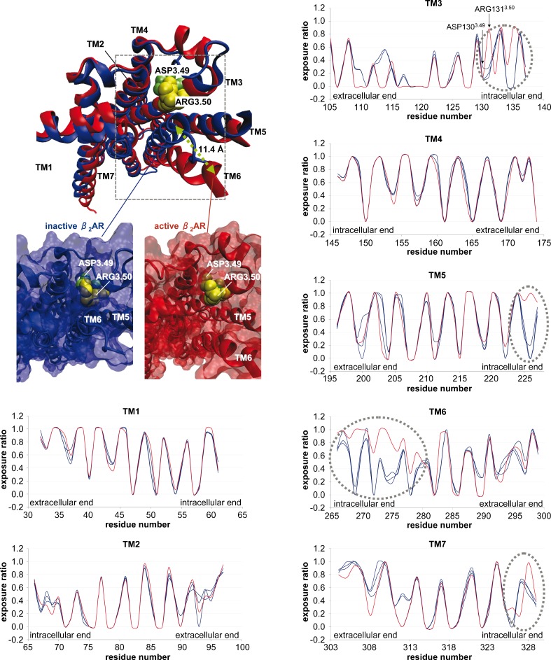
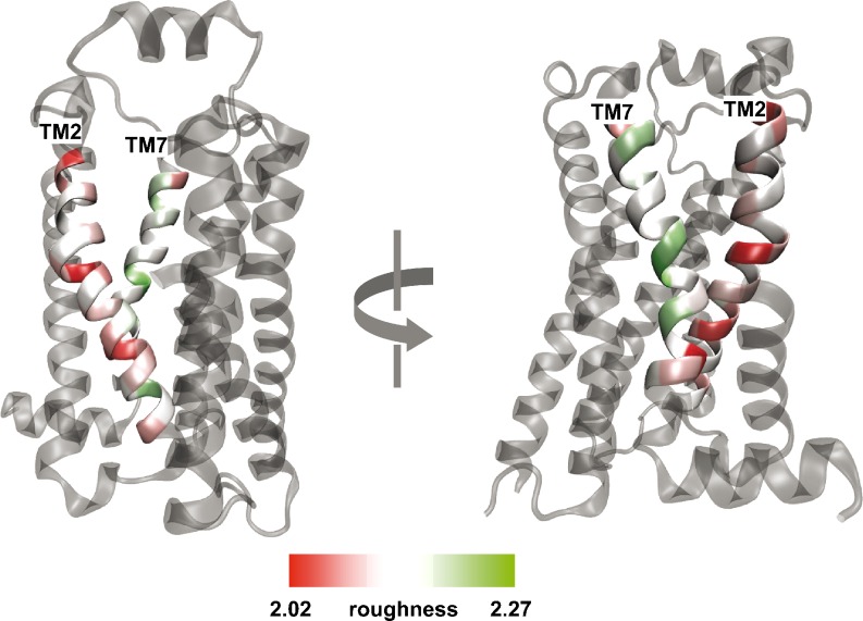
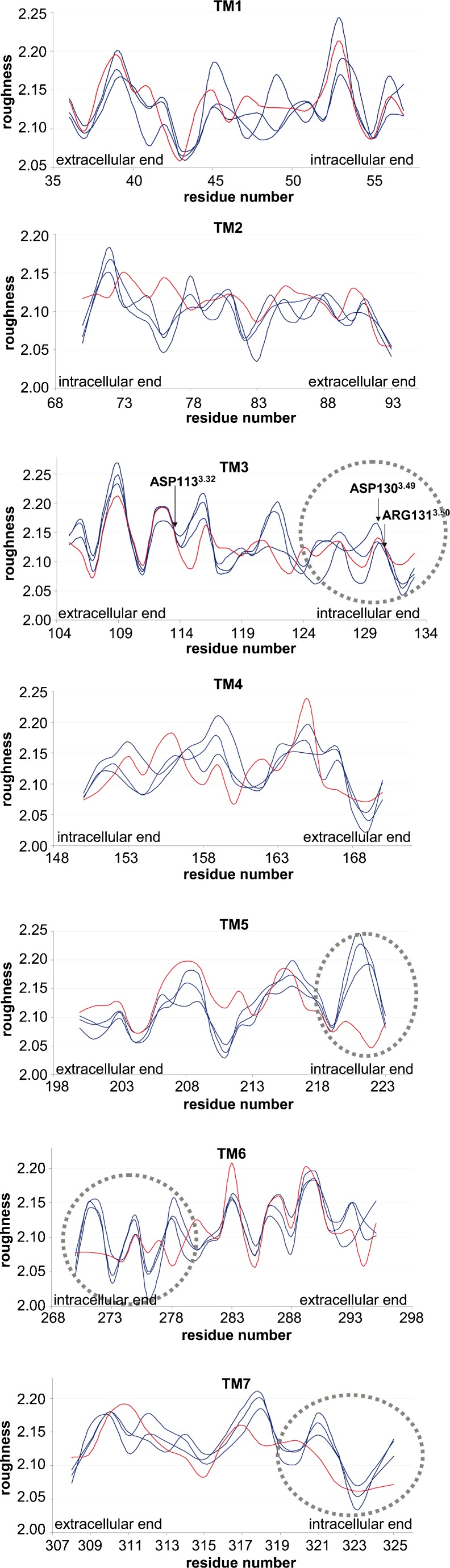
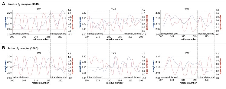
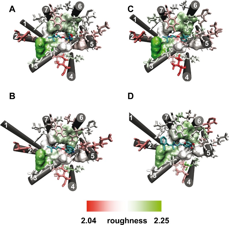
Protein surface roughness is a structural property associated with ligand-protein and protein-protein binding interfaces. In this work we apply for the first time the concept of surface roughness, expressed as the fractal dimension, to address structure and function of G protein-coupled receptors (GPCRs) which are an important group of drug targets. We calculate the exposure ratio and the fractal dimension for helix-forming residues of the β(2) adrenergic receptor (β(2)AR), a model system in GPCR studies, in different conformational states: in complex with agonist, antagonist and partial inverse agonists. We show that both exposure ratio and roughness exhibit periodicity which results from the helical structure of GPCRs. The pattern of roughness and exposure ratio of a protein patch depends on its environment: the residues most exposed to membrane are in general most rough whereas parts of receptors mediating interhelical contacts in a monomer or protein complex are much smoother. We also find that intracellular ends (TM3, TM5, TM6 and TM7) which are relevant for G protein binding and thus receptor signaling, are exposed but smooth. Mapping the values of residual fractal dimension onto receptor 3D structures makes it possible to conclude that the binding sites of orthosteric ligands as well as of cholesterol are characterized with significantly higher roughness than the average for the whole protein. In summary, our study suggests that identification of specific patterns of roughness could be a novel approach to spot possible binding sites which could serve as original drug targets for GPCRs modulation.
蛋白质表面粗糙度是与配体-蛋白质和蛋白质-蛋白质结合界面相关的结构特性。在这项工作中,我们首次将表面粗糙度的概念(表示为分形维数)应用于 G 蛋白偶联受体(GPCR)的结构和功能研究,GPCR 是一类重要的药物靶标。我们计算了β(2)肾上腺素能受体(β(2)AR)——GPCR 研究的模型体系——不同构象状态下形成螺旋的残基的暴露比和分形维数:与激动剂、拮抗剂和部分反向激动剂复合物。我们发现,暴露比和粗糙度都表现出周期性,这是由 GPCR 的螺旋结构引起的。蛋白质斑块的粗糙度和暴露比的模式取决于其环境:与膜最接近的残基通常最粗糙,而在单体或蛋白质复合物中介导螺旋间接触的受体部分则更光滑。我们还发现,与 G 蛋白结合以及受体信号转导相关的细胞内末端(TM3、TM5、TM6 和 TM7)是暴露的,但很光滑。将残差分形维数的值映射到受体 3D 结构上,可以得出结论,变构配体和胆固醇的结合位点的粗糙度明显高于整个蛋白质的平均水平。总之,我们的研究表明,识别特定的粗糙度模式可能是一种发现可能的结合位点的新方法,这些结合位点可以作为 GPCR 调节的原始药物靶标。