Department of Medical Imaging, Jinling Hospital, Clinical School of Medical College, Nanjing University, Nanjing, Jiangsu Province, China.
PLoS One. 2012;7(5):e37400. doi: 10.1371/journal.pone.0037400. Epub 2012 May 25.
Minimal hepatic encephalopathy (MHE) is a neuro-cognitive dysfunction characterized by impairment in attention, vigilance and integrative functions, while the sensorimotor function was often unaffected. Little is known, so far, about the exact neuro-pathophysiological mechanisms of aberrant cognition function in this disease.
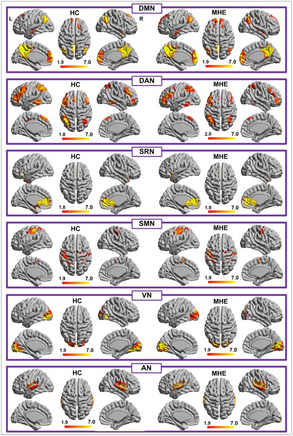
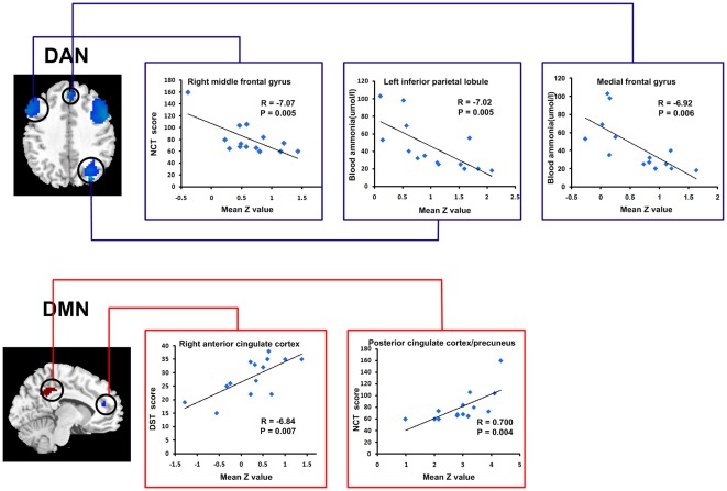
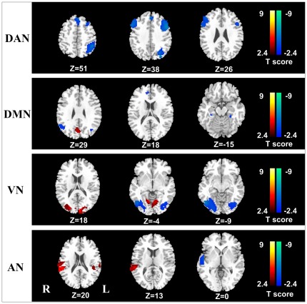
METHODOLOGY/PRINCIPAL FINDINGS: To investigate how the brain function is changed in MHE, we applied a resting-state fMRI approach with independent component analysis (ICA) to assess the differences of resting-state networks (RSNs) between MHE patients and healthy controls. Fourteen MHE patients and 14 age-and sex-matched healthy subjects underwent resting-state fMRI scans. ICA was used to identify six RSNs [dorsal attention network (DAN), default mode network (DMN), visual network (VN), auditory network (AN), sensorimotor network (SMN), self-referential network (SRN)] in each subject. Group maps of each RSN were compared between the MHE and healthy control groups. Pearson correlation analysis was performed between the RSNs functional connectivity (FC) and venous blood ammonia levels, and neuropsychological tests scores for all patients. Compared with the healthy controls, MHE patients showed significantly decreased FC in DAN, both decreased and increased FC in DMN, AN and VN. No significant differences were found in SRN and SMN between two groups. A relationship between FC and blood ammonia levels/neuropsychological tests scores were found in specific regions of RSNs, including middle and medial frontal gyrus, inferior parietal lobule, as well as anterior and posterior cingulate cortex/precuneus.
CONCLUSIONS/SIGNIFICANCE: MHE patients have selective impairments of RSNs intrinsic functional connectivity, with aberrant functional connectivity in DAN, DMN, VN, AN, and spared SMN and SRN. Our fMRI study might supply a novel way to understand the neuropathophysiological mechanism of cognition function changes in MHE.
轻微型肝性脑病(MHE)是一种以注意力、警觉和综合功能受损为特征的神经认知功能障碍,而感觉运动功能通常不受影响。迄今为止,对于这种疾病中异常认知功能的确切神经生理机制知之甚少。
方法/主要发现:为了研究 MHE 患者大脑功能的变化,我们应用静息态功能磁共振成像(rs-fMRI)结合独立成分分析(ICA)来评估 MHE 患者和健康对照组之间静息态网络(RSN)的差异。14 名 MHE 患者和 14 名年龄和性别匹配的健康受试者接受了 rs-fMRI 扫描。ICA 用于在每个受试者中识别六个 RSN[背侧注意网络(DAN)、默认模式网络(DMN)、视觉网络(VN)、听觉网络(AN)、感觉运动网络(SMN)、自我参照网络(SRN)]。比较 MHE 组和健康对照组之间每个 RSN 的组图。对所有患者的 RSN 功能连接(FC)与静脉血氨水平和神经心理学测试评分进行 Pearson 相关分析。与健康对照组相比,MHE 患者的 DAN 区 FC 明显降低,DMN、AN 和 VN 区 FC 降低或升高。两组间 SRN 和 SMN 无明显差异。在 RSN 的特定区域,包括中额叶和内侧额叶、下顶叶以及前后扣带回/楔前叶,发现 FC 与血液氨水平/神经心理学测试评分之间存在关系。
结论/意义:MHE 患者存在 RSN 固有功能连接的选择性损伤,DAN、DMN、VN、AN 的功能连接异常,而感觉运动网络和自我参照网络保持正常。我们的 fMRI 研究可能为理解 MHE 认知功能变化的神经病理生理机制提供新的方法。