• 文献检索
  • 文档翻译
  • 深度研究
  • 学术资讯
  • Suppr Zotero 插件Zotero 插件
  • 邀请有礼
  • 套餐&价格
  • 历史记录
应用&插件
Suppr Zotero 插件Zotero 插件浏览器插件Mac 客户端Windows 客户端微信小程序
定价
高级版会员购买积分包购买API积分包
服务
文献检索文档翻译深度研究API 文档MCP 服务
关于我们
关于 Suppr公司介绍联系我们用户协议隐私条款
关注我们

Suppr 超能文献

核心技术专利:CN118964589B侵权必究
粤ICP备2023148730 号-1Suppr @ 2026

文献检索

告别复杂PubMed语法,用中文像聊天一样搜索,搜遍4000万医学文献。AI智能推荐,让科研检索更轻松。

立即免费搜索

文件翻译

保留排版,准确专业,支持PDF/Word/PPT等文件格式,支持 12+语言互译。

免费翻译文档

深度研究

AI帮你快速写综述,25分钟生成高质量综述,智能提取关键信息,辅助科研写作。

立即免费体验

基于支持向量机学习算法的脑灰质容积测量对轻微型肝性脑病患者的识别。

Identification of patients with and without minimal hepatic encephalopathy based on gray matter volumetry using a support vector machine learning algorithm.

机构信息

College of Computer and Information Sciences, Fujian Agriculture and Forestry University, Fuzhou, 350002, China.

Department of Radiology, Fujian Medical University Union Hospital, Fuzhou, 350001, China.

出版信息

Sci Rep. 2020 Feb 12;10(1):2490. doi: 10.1038/s41598-020-59433-1.

DOI:10.1038/s41598-020-59433-1
PMID:32051514
原文链接:https://pmc.ncbi.nlm.nih.gov/articles/PMC7016173/
Abstract

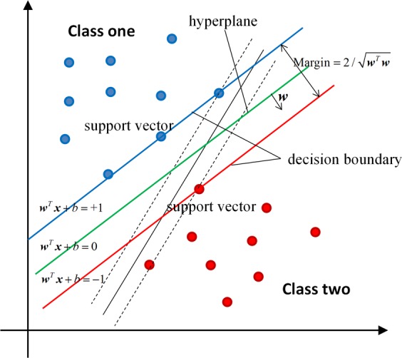
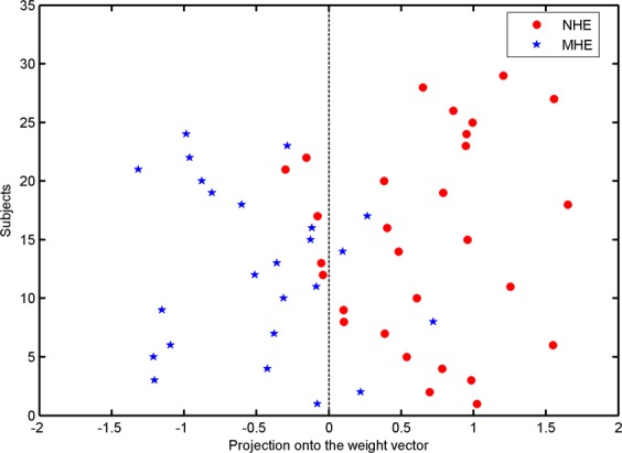
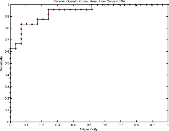
Minimal hepatic encephalopathy (MHE) is characterized by diffuse abnormalities in cerebral structure, such as reduced cortical thickness and altered brain parenchymal volume. This study tested the potential of gray matter (GM) volumetry to differentiate between cirrhotic patients with and without MHE using a support vector machine (SVM) learning method. High-resolution, T1-weighted magnetic resonance images were acquired from 24 cirrhotic patients with MHE and 29 cirrhotic patients without MHE (NHE). Voxel-based morphometry was conducted to evaluate the GM volume (GMV) for each subject. An SVM classifier was employed to explore the ability of the GMV measurement to diagnose MHE, and the leave-one-out cross-validation method was used to assess classification accuracy. The SVM algorithm based on GM volumetry achieved a classification accuracy of 83.02%, with a sensitivity of 83.33% and a specificity of 82.76%. The majority of the most discriminative GMVs were located in the bilateral frontal lobe, bilateral lentiform nucleus, bilateral thalamus, bilateral sensorimotor areas, bilateral visual regions, bilateral temporal lobe, bilateral cerebellum, left inferior parietal lobe, and right precuneus/posterior cingulate gyrus. Our results suggest that SVM analysis based on GM volumetry has the potential to help diagnose MHE in cirrhotic patients.

摘要

轻微型肝性脑病(MHE)的特征是脑结构弥漫性异常,如皮质厚度降低和脑实质体积改变。本研究采用支持向量机(SVM)学习方法,测试了基于灰质(GM)体积测量区分伴或不伴 MHE 的肝硬化患者的潜力。从 24 例 MHE 肝硬化患者和 29 例非 MHE(NHE)肝硬化患者中采集了高分辨率 T1 加权磁共振图像。对每个受试者进行基于体素的形态测量,以评估 GM 体积(GMV)。采用 SVM 分类器探讨 GMV 测量对 MHE 的诊断能力,采用留一法交叉验证法评估分类准确性。基于 GM 容积的 SVM 算法实现了 83.02%的分类准确率,敏感性为 83.33%,特异性为 82.76%。大多数最具判别力的 GMVs 位于双侧额叶、双侧豆状核、双侧丘脑、双侧感觉运动区、双侧视觉区域、双侧颞叶、双侧小脑、左侧顶下小叶和右侧楔前叶/后扣带回。我们的结果表明,基于 GM 体积的 SVM 分析有可能有助于诊断肝硬化患者的 MHE。

https://cdn.ncbi.nlm.nih.gov/pmc/blobs/9d1f/7016173/144471c53796/41598_2020_59433_Fig4_HTML.jpg
https://cdn.ncbi.nlm.nih.gov/pmc/blobs/9d1f/7016173/6c485b08c60b/41598_2020_59433_Fig1_HTML.jpg
https://cdn.ncbi.nlm.nih.gov/pmc/blobs/9d1f/7016173/53f5a019c148/41598_2020_59433_Fig2_HTML.jpg
https://cdn.ncbi.nlm.nih.gov/pmc/blobs/9d1f/7016173/d3c051ff0742/41598_2020_59433_Fig3_HTML.jpg
https://cdn.ncbi.nlm.nih.gov/pmc/blobs/9d1f/7016173/144471c53796/41598_2020_59433_Fig4_HTML.jpg

相似文献

1
Identification of patients with and without minimal hepatic encephalopathy based on gray matter volumetry using a support vector machine learning algorithm.基于支持向量机学习算法的脑灰质容积测量对轻微型肝性脑病患者的识别。
Sci Rep. 2020 Feb 12;10(1):2490. doi: 10.1038/s41598-020-59433-1.
2
Identifying minimal hepatic encephalopathy in cirrhotic patients by measuring spontaneous brain activity.通过测量自发性脑活动来识别肝硬化患者的轻微肝性脑病。
Metab Brain Dis. 2016 Aug;31(4):761-9. doi: 10.1007/s11011-016-9799-9. Epub 2016 Feb 17.
3
Machine Learning Classification of Cirrhotic Patients with and without Minimal Hepatic Encephalopathy Based on Regional Homogeneity of Intrinsic Brain Activity.基于脑内固有活动区域同质性的机器学习对有无轻微肝性脑病肝硬化患者的分类
PLoS One. 2016 Mar 15;11(3):e0151263. doi: 10.1371/journal.pone.0151263. eCollection 2016.
4
Hippocampal atrophy and functional connectivity disruption in cirrhotic patients with minimal hepatic encephalopathy.肝硬化伴轻微肝性脑病患者的海马萎缩和功能连接中断。
Metab Brain Dis. 2019 Dec;34(6):1519-1529. doi: 10.1007/s11011-019-00457-6. Epub 2019 Jul 30.
5
Identification of minimal hepatic encephalopathy based on dynamic functional connectivity.基于动态功能连接的轻微型肝性脑病的识别。
Brain Imaging Behav. 2021 Oct;15(5):2637-2645. doi: 10.1007/s11682-021-00468-x. Epub 2021 Mar 23.
6
Altered dynamic functional connectivity in the default mode network in patients with cirrhosis and minimal hepatic encephalopathy.肝硬化合并轻微肝性脑病患者默认模式网络中动态功能连接的改变。
Neuroradiology. 2017 Sep;59(9):905-914. doi: 10.1007/s00234-017-1881-4. Epub 2017 Jul 13.
7
Cerebral blood flow measured by arterial-spin labeling MRI: a useful biomarker for characterization of minimal hepatic encephalopathy in patients with cirrhosis.动脉自旋标记 MRI 测量脑血流:肝硬化患者轻微肝性脑病特征描述的有用生物标志物。
Eur J Radiol. 2013 Nov;82(11):1981-8. doi: 10.1016/j.ejrad.2013.06.002. Epub 2013 Jul 9.
8
Reduced resting state connectivity and gray matter volume correlate with cognitive impairment in minimal hepatic encephalopathy.静息态连接性降低和灰质体积减少与轻微肝性脑病的认知障碍相关。
PLoS One. 2017 Oct 12;12(10):e0186463. doi: 10.1371/journal.pone.0186463. eCollection 2017.
9
Abnormal spontaneous brain activity in minimal hepatic encephalopathy: resting-state fMRI study.轻微肝性脑病患者的异常自发性脑活动:静息态功能磁共振成像研究
Diagn Interv Radiol. 2016 Mar-Apr;22(2):196-200. doi: 10.5152/dir.2015.15208.
10
Disrupted thalamic resting-state functional connectivity in patients with minimal hepatic encephalopathy.轻微型肝性脑病患者丘脑静息态功能连接中断。
Eur J Radiol. 2013 May;82(5):850-6. doi: 10.1016/j.ejrad.2012.12.016. Epub 2013 Jan 17.

引用本文的文献

1
Machine learning techniques in hepatic encephalopathy: a scoping review.肝性脑病中的机器学习技术:一项范围综述
BMC Med Inform Decis Mak. 2025 Sep 1;25(1):323. doi: 10.1186/s12911-025-03168-4.
2
Minimal hepatic encephalopathy: Characteristics and comparison of the main diagnostic modalities.轻微肝性脑病:主要诊断方法的特点及比较
Clin Exp Hepatol. 2024 Dec;10(4):218-226. doi: 10.5114/ceh.2024.145438. Epub 2024 Dec 2.
3
Normative Modeling Reveals Age-Atypical Cortical Thickness Differences Between Hepatic Steatosis and Fibrosis in Non-Alcoholic Fatty Liver Disease.

本文引用的文献

1
Resting-state functional connectivity abnormalities correlate with psychometric hepatic encephalopathy score in cirrhosis.静息态功能连接异常与肝硬化患者的心理测量肝性脑病评分相关。
Eur J Radiol. 2015 Nov;84(11):2287-95. doi: 10.1016/j.ejrad.2015.08.005. Epub 2015 Aug 18.
2
Cortical signature of patients with HBV-related cirrhosis without overt hepatic encephalopathy: a morphometric analysis.乙肝相关肝硬化且无明显肝性脑病患者的皮质特征:一项形态学分析
Front Neuroanat. 2015 Jun 8;9:82. doi: 10.3389/fnana.2015.00082. eCollection 2015.
3
Hepatic encephalopathy in chronic liver disease: 2014 Practice Guideline by the American Association for the Study of Liver Diseases and the European Association for the Study of the Liver.
规范建模揭示了非酒精性脂肪性肝病中肝脂肪变性和肝纤维化之间的年龄非典型皮质厚度差异。
Brain Behav. 2025 Apr;15(4):e70466. doi: 10.1002/brb3.70466.
4
Changes in dynamic and static brain fluctuation distinguish minimal hepatic encephalopathy and cirrhosis patients and predict the severity of liver damage.动态和静态脑波动的变化可区分轻微肝性脑病患者和肝硬化患者,并预测肝损伤的严重程度。
Front Neurosci. 2023 Mar 28;17:1077808. doi: 10.3389/fnins.2023.1077808. eCollection 2023.
5
The Link between Gut Microbiota and Hepatic Encephalopathy.肠道微生物群与肝性脑病的关系。
Int J Mol Sci. 2022 Aug 12;23(16):8999. doi: 10.3390/ijms23168999.
6
Artificial Intelligence and Its Application to Minimal Hepatic Encephalopathy Diagnosis.人工智能及其在轻微肝性脑病诊断中的应用。
J Pers Med. 2021 Oct 26;11(11):1090. doi: 10.3390/jpm11111090.
7
Reduced Cortical Complexity in Cirrhotic Patients with Minimal Hepatic Encephalopathy.肝硬化伴轻微肝性脑病患者皮质复杂度降低。
Neural Plast. 2020 Mar 18;2020:7364649. doi: 10.1155/2020/7364649. eCollection 2020.
慢性肝病中的肝性脑病:美国肝病研究协会和欧洲肝脏研究协会2014年实践指南
Hepatology. 2014 Aug;60(2):715-35. doi: 10.1002/hep.27210. Epub 2014 Jul 8.
4
Multivariate pattern analysis of DTI reveals differential white matter in individuals with obsessive-compulsive disorder.弥散张量成像的多变量模式分析揭示了强迫症患者白质的差异。
Hum Brain Mapp. 2014 Jun;35(6):2643-51. doi: 10.1002/hbm.22357. Epub 2013 Sep 18.
5
PRoNTo: pattern recognition for neuroimaging toolbox.PRoNTo:神经影像学工具包的模式识别。
Neuroinformatics. 2013 Jul;11(3):319-37. doi: 10.1007/s12021-013-9178-1.
6
Brain dysfunction primarily related to previous overt hepatic encephalopathy compared with minimal hepatic encephalopathy: resting-state functional MR imaging demonstration.与轻微型肝性脑病相比,主要与先前显性肝性脑病相关的脑功能障碍:静息状态功能磁共振成像的显示。
Radiology. 2013 Jan;266(1):261-70. doi: 10.1148/radiol.12120026. Epub 2012 Oct 9.
7
The effect of hepatic encephalopathy, hepatic failure, and portosystemic shunt on brain volume of cirrhotic patients: a voxel-based morphometry study.肝性脑病、肝衰竭和门体分流对肝硬化患者脑容量的影响:基于体素的形态计量学研究。
PLoS One. 2012;7(8):e42824. doi: 10.1371/journal.pone.0042824. Epub 2012 Aug 13.
8
Selective impairments of resting-state networks in minimal hepatic encephalopathy.轻微型肝性脑病的静息态网络选择性损伤。
PLoS One. 2012;7(5):e37400. doi: 10.1371/journal.pone.0037400. Epub 2012 May 25.
9
Regional reduction in gray and white matter volume in brains of cirrhotic patients: voxel-based analysis of MRI.肝硬化患者脑灰质和白质体积的区域性减少:MRI 的体素基分析。
Metab Brain Dis. 2012 Dec;27(4):551-7. doi: 10.1007/s11011-012-9314-x. Epub 2012 May 18.
10
Focal cortical damage parallels cognitive impairment in minimal hepatic encephalopathy.轻微肝性脑病的皮质局部损伤与认知障碍平行。
Neuroimage. 2012 Jul 16;61(4):1165-75. doi: 10.1016/j.neuroimage.2012.03.041. Epub 2012 Mar 21.