• 文献检索
  • 文档翻译
  • 深度研究
  • 学术资讯
  • Suppr Zotero 插件Zotero 插件
  • 邀请有礼
  • 套餐&价格
  • 历史记录
应用&插件
Suppr Zotero 插件Zotero 插件浏览器插件Mac 客户端Windows 客户端微信小程序
定价
高级版会员购买积分包购买API积分包
服务
文献检索文档翻译深度研究API 文档MCP 服务
关于我们
关于 Suppr公司介绍联系我们用户协议隐私条款
关注我们

Suppr 超能文献

核心技术专利:CN118964589B侵权必究
粤ICP备2023148730 号-1Suppr @ 2026

文献检索

告别复杂PubMed语法,用中文像聊天一样搜索,搜遍4000万医学文献。AI智能推荐,让科研检索更轻松。

立即免费搜索

文件翻译

保留排版,准确专业,支持PDF/Word/PPT等文件格式,支持 12+语言互译。

免费翻译文档

深度研究

AI帮你快速写综述,25分钟生成高质量综述,智能提取关键信息,辅助科研写作。

立即免费体验

通过基于微泡的超声造影评估硬皮病患者的肾灌注。

Renal perfusion in scleroderma patients assessed by microbubble-based contrast-enhanced ultrasound.

作者信息

Kleinert Stefan, Roll Petra, Baumgaertner Christian, Himsel Andrea, Mueller Adelheid, Fleck Martin, Feuchtenberger Martin, Jenett Manfred, Tony Hans-Peter

机构信息

University Hospital of Wuerzburg, Rheumatology/Clinical Immunology, Oberduerrbacherstr. 6, D-97080 Wuerzburg, Germany.

出版信息

Open Rheumatol J. 2012;6:50-3. doi: 10.2174/1874312901206010050. Epub 2012 May 30.

DOI:10.2174/1874312901206010050
PMID:22670165
原文链接:https://pmc.ncbi.nlm.nih.gov/articles/PMC3367302/
Abstract

OBJECTIVES

Renal damage is common in scleroderma. It can occur acutely or chronically. Renal reserve might already be impaired before it can be detected by laboratory findings. Microbubble-based contrast-enhanced ultrasound has been demonstrated to improve blood perfusion imaging in organs. Therefore, we conducted a study to assess renal perfusion in scleroderma patients utilizing this novel technique.

MATERIALS AND METHODOLOGY

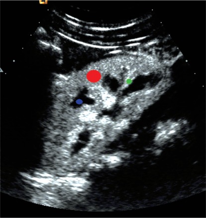
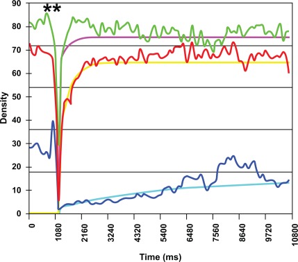
Microbubble-based contrast agent was infused and destroyed by using high mechanical index by Siemens Sequoia (curved array, 4.5 MHz). Replenishment was recorded for 8 seconds. Regions of interests (ROI) were analyzed in renal parenchyma, interlobular artery and renal pyramid with quantitative contrast software (CUSQ 1.4, Siemens Acuson, Mountain View, California). Time to maximal Enhancement (TmE), maximal enhancement (mE) and maximal enhancement relative to maximal enhancement of the interlobular artery (mE%A) were calculated for different ROIs.

RESULTS

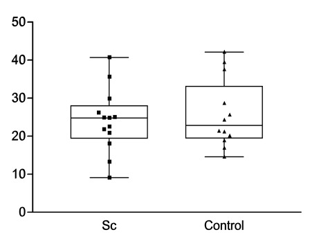
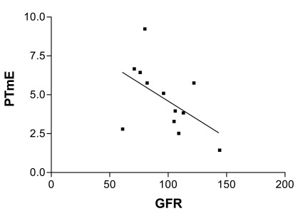
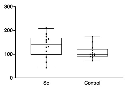
There was a linear correlation between the time to maximal enhancement in the parenchyma and the glomerular filtration rate. However, the other parameters did not reveal significant differences between scleroderma patients and healthy controls.

CONCLUSION

Renal perfusion of scleroderma patients including the glomerular filtration rate can be assessed using microbubble-based contrast media.

摘要

目的

肾脏损害在硬皮病中很常见。它可急性或慢性发生。在实验室检查结果能够检测到之前,肾脏储备功能可能就已经受损。基于微泡的超声造影已被证明可改善器官的血流灌注成像。因此,我们开展了一项研究,利用这项新技术评估硬皮病患者的肾脏灌注情况。

材料与方法

使用西门子Sequoia(弯阵探头,4.5MHz)通过高机械指数注入并破坏基于微泡的造影剂。记录8秒的再充盈情况。使用定量造影软件(CUSQ 1.4,西门子Acuson,加利福尼亚州山景城)对肾实质、小叶间动脉和肾锥体中的感兴趣区域(ROI)进行分析。计算不同ROI的达峰时间(TmE)、最大增强(mE)以及相对于小叶间动脉最大增强的最大增强百分比(mE%A)。

结果

肾实质的达峰时间与肾小球滤过率之间存在线性相关性。然而,硬皮病患者与健康对照之间的其他参数未显示出显著差异。

结论

使用基于微泡的造影剂可以评估硬皮病患者的肾脏灌注情况,包括肾小球滤过率。

https://cdn.ncbi.nlm.nih.gov/pmc/blobs/b2e6/3367302/b9b4128f78df/TORJ-6-50_F5.jpg
https://cdn.ncbi.nlm.nih.gov/pmc/blobs/b2e6/3367302/bd0c1fbb8e47/TORJ-6-50_F1.jpg
https://cdn.ncbi.nlm.nih.gov/pmc/blobs/b2e6/3367302/2a699f1705ff/TORJ-6-50_F2.jpg
https://cdn.ncbi.nlm.nih.gov/pmc/blobs/b2e6/3367302/d5e2b1cc9ad6/TORJ-6-50_F3.jpg
https://cdn.ncbi.nlm.nih.gov/pmc/blobs/b2e6/3367302/e768f2e8c4fb/TORJ-6-50_F4.jpg
https://cdn.ncbi.nlm.nih.gov/pmc/blobs/b2e6/3367302/b9b4128f78df/TORJ-6-50_F5.jpg

相似文献

1
Renal perfusion in scleroderma patients assessed by microbubble-based contrast-enhanced ultrasound.通过基于微泡的超声造影评估硬皮病患者的肾灌注。
Open Rheumatol J. 2012;6:50-3. doi: 10.2174/1874312901206010050. Epub 2012 May 30.
2
Evaluation of Acoustic Radiation Force Impulse (ARFI) imaging and contrast-enhanced ultrasound in renal tumors of unknown etiology in comparison to histological findings.评估声辐射力脉冲(ARFI)成像和超声造影在与组织学发现相比对不明原因肾肿瘤的诊断价值。
Clin Hemorheol Microcirc. 2009;43(1-2):95-107. doi: 10.3233/CH-2009-1224.
3
Italian Society of Cardiovascular Echography (SIEC) Consensus Conference on the state of the art of contrast echocardiography.意大利心血管超声学会(SIEC)关于超声造影技术现状的共识会议
Ital Heart J. 2004 Apr;5(4):309-34.
4
Renal parenchyma perfusion spectrum and resistive index (RI) in ultrasound examinations with contrast medium in the early period after kidney transplantation.肾移植术后早期超声造影检查中肾实质灌注频谱及阻力指数(RI)
Transplant Proc. 2009 Oct;41(8):3024-7. doi: 10.1016/j.transproceed.2009.08.023.
5
Correlation between estimates of tumor perfusion from microbubble contrast-enhanced sonography and dynamic contrast-enhanced magnetic resonance imaging.微泡对比增强超声检查与动态对比增强磁共振成像评估肿瘤灌注之间的相关性
J Ultrasound Med. 2006 Apr;25(4):487-97. doi: 10.7863/jum.2006.25.4.487.
6
[Renal cell cancer does not show a typical perfusion pattern in contrast-enhanced ultrasound].与正常组织相比,肾细胞癌在超声造影中不显示典型的灌注模式。
Ultraschall Med. 2009 Feb;30(1):58-63. doi: 10.1055/s-2008-1027189.
7
Renal perfusion parameters measured by contrast-enhanced ultrasound in healthy dogs demonstrate a wide range of variability in the long-term.通过超声造影测量的健康犬的肾灌注参数在长期内表现出广泛的变异性。
Vet Radiol Ultrasound. 2019 Mar;60(2):201-209. doi: 10.1111/vru.12690. Epub 2018 Oct 1.
8
CT perfusion of renal cell carcinoma: impact of volume coverage on quantitative analysis.CT 灌注成像在肾细胞癌中的应用:容积覆盖范围对定量分析的影响。
Invest Radiol. 2012 Jan;47(1):33-40. doi: 10.1097/RLI.0b013e31822598c3.
9
Repeatability of the "flash-replenishment" method in contrast-enhanced ultrasound for the quantitative assessment of hepatic microvascular perfusion.超声造影“快速补充”法对肝脏微血管灌注进行定量评估的可重复性
Braz J Med Biol Res. 2018;51(4):e7058. doi: 10.1590/1414-431x20177058. Epub 2018 Feb 26.
10
Comparison between echo contrast agent-specific imaging modes and perfusion-weighted magnetic resonance imaging for the assessment of brain perfusion.用于评估脑灌注的超声造影剂特异性成像模式与灌注加权磁共振成像之间的比较。
Stroke. 2002 Oct;33(10):2433-7. doi: 10.1161/01.str.0000032246.85531.8e.

引用本文的文献

1
Usefulness of Contrast-enhanced Ultrasound in the Evaluation of Chronic Kidney Disease.超声造影在慢性肾脏病评估中的应用价值
Curr Med Imaging. 2021;17(8):1003-1009. doi: 10.2174/1573405617666210127101926.
2
Clinical significance of contrast-enhanced ultrasound in chronic kidney disease: a pilot study.超声造影在慢性肾脏病中的临床意义:一项初步研究。
J Ultrasound. 2019 Dec;22(4):453-460. doi: 10.1007/s40477-019-00409-x. Epub 2019 Oct 12.
3
Effects of high-fat diet and losartan on renal cortical blood flow using contrast ultrasound imaging.

本文引用的文献

1
Perfusion of kidney graft pyramids and cortex in contrast-enhanced ultrasonography in the determination of the cause of delayed graft function.肾移植锥体和皮质灌注的对比增强超声检查在确定移植肾功能延迟原因中的应用
Ann Transplant. 2011 Jan-Mar;16(1):48-53.
2
Early postoperative ultrasound of kidney transplants: evaluation of contrast medium dynamics using time-intensity curves.肾移植术后早期超声检查:使用时间-强度曲线评估造影剂动力学
Rofo. 2004 Apr;176(4):472-7. doi: 10.1055/s-2004-812992.
3
Blood flow quantification with contrast-enhanced US: "entrance in the section" phenomenon--phantom and rabbit study.
高脂饮食和氯沙坦对应用对比超声成像的肾皮质血流的影响。
Am J Physiol Renal Physiol. 2013 Nov 1;305(9):F1343-51. doi: 10.1152/ajprenal.00326.2013. Epub 2013 Sep 18.
超声造影血流定量:“进入切面”现象——体模与兔实验研究
Radiology. 2003 Aug;228(2):473-9. doi: 10.1148/radiol.2282020699. Epub 2003 Jun 11.
4
Renal functional reserve is impaired in patients with systemic sclerosis without clinical signs of kidney involvement.在无肾脏受累临床体征的系统性硬化症患者中,肾功能储备受损。
Ann Rheum Dis. 2002 Aug;61(8):682-6. doi: 10.1136/ard.61.8.682.
5
A more accurate method to estimate glomerular filtration rate from serum creatinine: a new prediction equation. Modification of Diet in Renal Disease Study Group.一种根据血清肌酐估算肾小球滤过率的更准确方法:一个新的预测方程。肾脏病饮食改良研究组。
Ann Intern Med. 1999 Mar 16;130(6):461-70. doi: 10.7326/0003-4819-130-6-199903160-00002.
6
Survival with scleroderma. II. A life-table analysis of clinical and demographic factors in 358 male U.S. veteran patients.
J Chronic Dis. 1973 Oct;26(10):647-60. doi: 10.1016/0021-9681(73)90054-4.
7
Survival with systemic sclerosis (scleroderma). A life-table analysis of clinical and demographic factors in 309 patients.
Ann Intern Med. 1971 Sep;75(3):369-76. doi: 10.7326/0003-4819-75-3-369.