Neuroscience and Psychiatry Unit, University of Manchester and Manchester Academic Health Sciences Centre, Manchester, United Kingdom.
Biol Psychiatry. 2012 Oct 1;72(7):604-11. doi: 10.1016/j.biopsych.2012.04.031. Epub 2012 Jun 6.
Vulnerability to relapse persists after remission of an acute episode of major depressive disorder. This has been attributed to abnormal biases in the processing of emotional stimuli in limbic circuits. However, neuroimaging studies have not so far revealed consistent evidence of abnormal responses to emotional stimuli in limbic structures, such as the amygdala, in remitted depression. This suggests the problem might lie in the integrated functioning of emotion processing circuits.
We recruited 22 unmedicated patients in remission from major depressive disorder (rMDD) and 21 age-matched healthy control subjects. Functional magnetic resonance imaging was performed during a face emotion processing task. Dynamic causal modeling was used with Bayesian model selection to determine the most likely brain networks and valence-specific modulation of connectivity in healthy control subjects and rMDD.
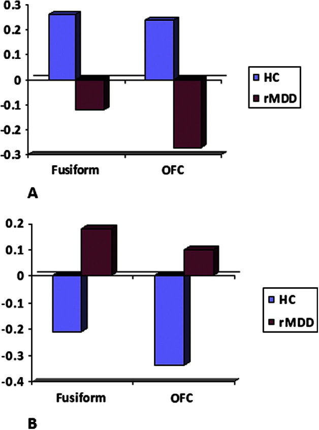
In healthy volunteers, sad faces modulated bi-directional connections between amygdala and orbitofrontal cortex and between fusiform gyrus and orbitofrontal cortex. Happy faces modulated unidirectional connections from fusiform gyrus to orbitofrontal cortex. In rMDD, the opposite pattern was observed, with evidence of happy faces modulating bidirectional frontotemporal connections and sad faces modulating unidirectional fusiform-orbitofrontal connections.
Participants with rMDD have abnormal modulation of frontotemporal effective connectivity in response to happy and sad face emotions, despite normal activations within each region. Specifically, processing of mood incongruent happy information was associated with a more richly modulated frontotemporal brain network, whereas mood congruent sad information was associated with less network modulation. This supports a hypothesis of dysfunction within cortico-limbic connections in individuals vulnerable to depression.
重度抑郁症急性发作缓解后,仍存在复发的脆弱性。这归因于边缘回路中情绪刺激处理的异常偏差。然而,神经影像学研究尚未揭示出在缓解期抑郁症中,边缘结构(如杏仁核)对情绪刺激反应异常的一致证据。这表明问题可能在于情绪处理回路的综合功能。
我们招募了 22 名未接受药物治疗的缓解期重度抑郁症患者(rMDD)和 21 名年龄匹配的健康对照者。在进行面部情绪处理任务时进行了功能磁共振成像。使用贝叶斯模型选择的动态因果建模来确定健康对照组和 rMDD 中最可能的大脑网络和与效价特异性连接的调制。
在健康志愿者中,悲伤的面孔调节了杏仁核和眶额皮质之间以及梭状回和眶额皮质之间的双向连接。快乐的面孔调节了从梭状回到眶额皮质的单向连接。在 rMDD 中,观察到相反的模式,即有证据表明快乐的面孔调节了额颞叶的双向连接,悲伤的面孔调节了梭状回-眶额皮质的单向连接。
尽管每个区域的激活正常,但 rMDD 参与者对快乐和悲伤面孔情绪的额颞叶有效连接存在异常调节。具体而言,与情绪不一致的快乐信息的处理与更丰富的额颞叶网络调节有关,而与情绪一致的悲伤信息的处理与较少的网络调节有关。这支持了易患抑郁症个体皮质-边缘连接功能障碍的假说。