Department of Orthopedics, The Second Affiliated Hospital of Soochow University, Suzhou, China.
BMC Musculoskelet Disord. 2012 Jun 8;13:94. doi: 10.1186/1471-2474-13-94.
It has been indicated that moderate or high dose of X-irradiation could delay fracture union and cause osteoradionecrosis, in part, mediated by its effect on proliferation and differentiation of osteoblasts. However, whether low dose irradiation (LDI) has similar roles on osteoblasts is still unknown. In this study, we investigated whether and to what extent LDI could affect the proliferation, differentiation and mineralization of osteoblasts in vitro.
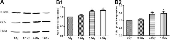
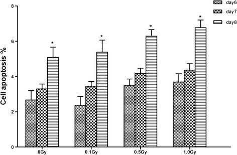
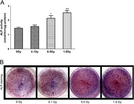
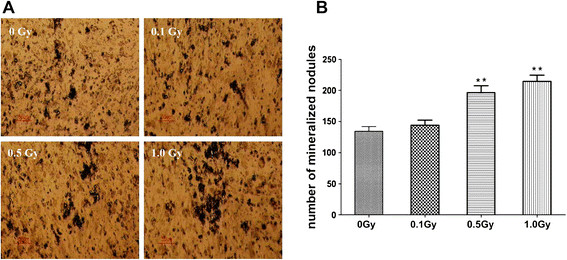
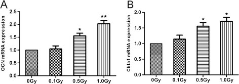
The MC3T3-E1 cells were exposed to single dose of X-irradiation with 0, 0.1, 0.5, 1.0 Gy respectively. Cell proliferation, apoptosis, alkaline phosphatase (ALP) activity, and mineralization was evaluated by methylthiazoletetrazolium (MTT) and bromodeoxyuridine (BrdU) assay, flow cytometry, ALP viability kit and von Kossa staining, respectively. Osteocalcin (OCN) and core-binding factor α1 (Cbfα1) expressions were measured by real time-PCR and western blot, respectively.
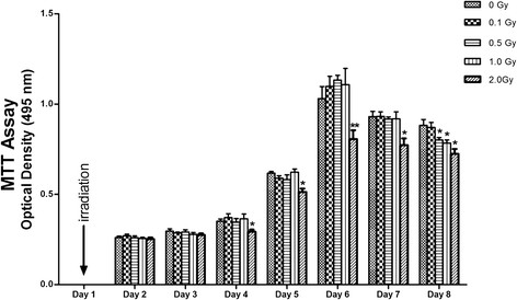
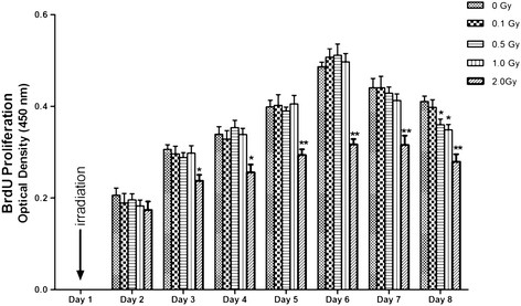
The proliferation of the cells exposed to 2.0 Gy was significantly lower than those exposed to ≤1.0 Gy (p < 0.05) from Day 4 to Day 8, measured by MTT assay and BrdU incorporation. For cells exposed to ≤1.0 Gy, increasing dosages of X-irradiation had no significant effect on cell proliferation and apoptosis. Importantly, LDI of 0.5 and 1 Gy increased ALP activities and mineralized nodules of MC3T3-E1 cells. In addition, mRNA and protein expressions of OCN and Cbfα1 were also markedly increased after treatment with LDI at 0.5 and 1 Gy.
LDI have different effects on proliferation and differentiation of osteoblasts from those of high dose of X-irradiation, which might suggest that LDI could lead to promotion of fracture healing through enhancing the differentiation and mineralization of osteoblasts.
有研究表明,中高剂量的 X 射线照射会延迟骨折愈合,并导致骨放射性坏死,部分原因是其对成骨细胞增殖和分化的影响。然而,低剂量照射(LDI)是否对成骨细胞有类似的作用尚不清楚。在这项研究中,我们研究了 LDI 是否以及在何种程度上会影响体外成骨细胞的增殖、分化和矿化。
用 0、0.1、0.5、1.0 Gy 的 X 射线分别照射 MC3T3-E1 细胞。通过甲基噻唑基四唑(MTT)和溴脱氧尿苷(BrdU)检测、流式细胞术、碱性磷酸酶(ALP)活力试剂盒和 von Kossa 染色分别评估细胞增殖、细胞凋亡、碱性磷酸酶(ALP)活性和矿化。通过实时 PCR 和 Western blot 分别测量骨钙素(OCN)和核心结合因子α1(Cbfα1)的表达。
与≤1.0 Gy 组相比,2.0 Gy 组细胞在第 4 天至第 8 天的 MTT 检测和 BrdU 掺入试验中增殖显著降低(p<0.05)。对于≤1.0 Gy 组,X 射线剂量的增加对细胞增殖和凋亡没有显著影响。重要的是,0.5 和 1 Gy 的 LDI 增加了 MC3T3-E1 细胞的 ALP 活性和矿化结节。此外,在 0.5 和 1 Gy 的 LDI 处理后,OCN 和 Cbfα1 的 mRNA 和蛋白表达也显著增加。
LDI 对成骨细胞增殖和分化的影响与高剂量 X 射线不同,这可能表明 LDI 通过增强成骨细胞的分化和矿化来促进骨折愈合。