Zhang Jian, Wang Ziyang, Wu Anqing, Nie Jing, Pei Hailong, Hu Wentao, Wang Bing, Shang Peng, Li Bingyan, Zhou Guangming
School of Radiation Medicine and Protection, Medical College of Soochow University, 199 Renai Road, Suzhou 215123, China.
Collaborative Innovation Center of Radiation Medicine of Jiangsu Higher Education Institutions, 199 Renai Road, Suzhou 215123, China.
J Radiat Res. 2017 Nov 1;58(6):791-802. doi: 10.1093/jrr/rrx026.
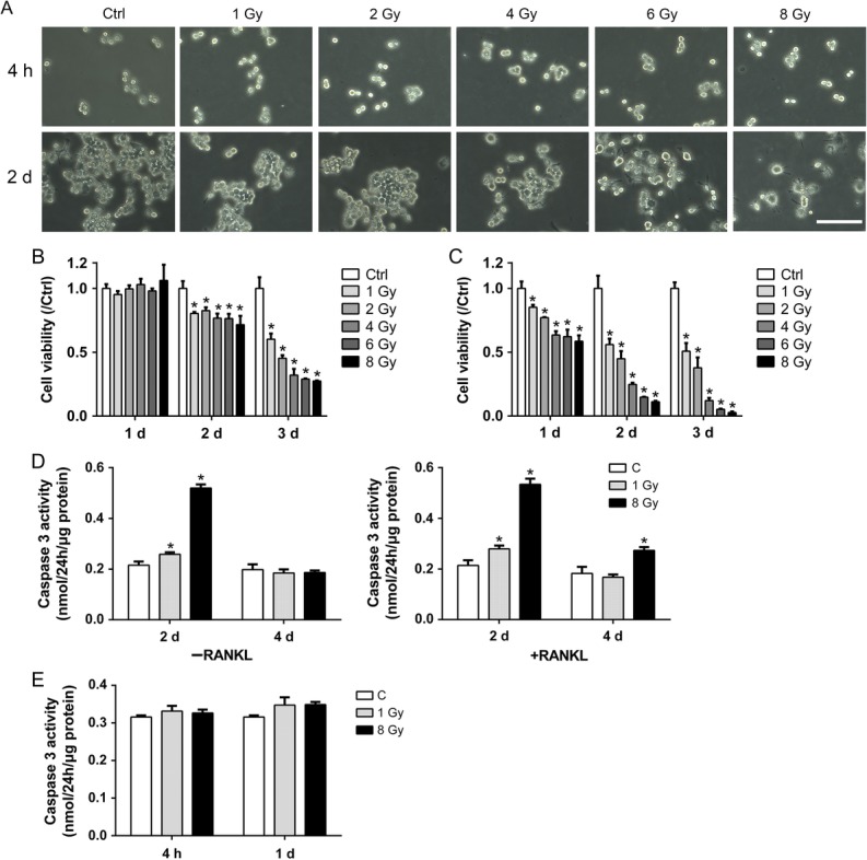
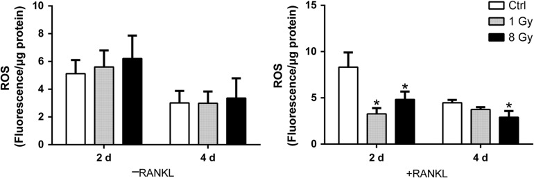
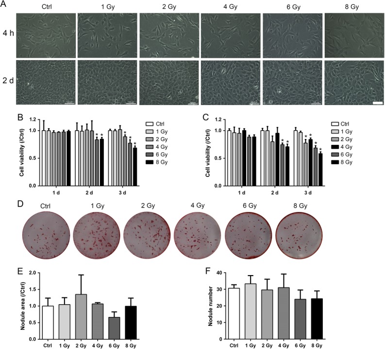
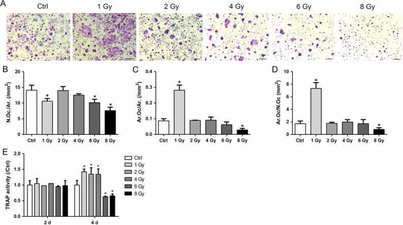
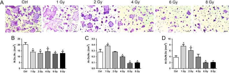
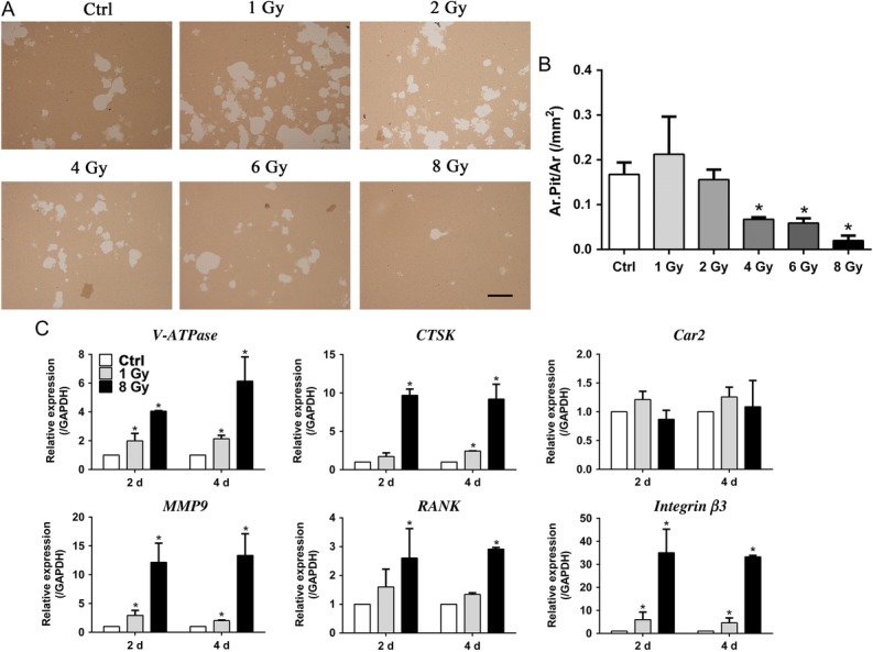
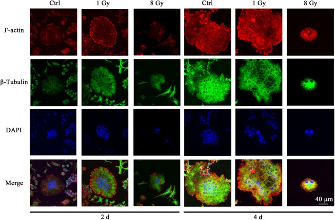
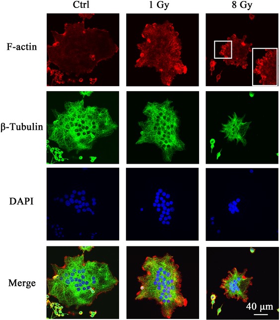
Radiation-induced bone loss is a potential health concern for cancer patients undergoing radiotherapy. Enhanced bone resorption by osteoclasts and decreased bone formation by osteoblasts were thought to be the main reasons. In this study, we showed that both pre-differentiating and differentiating osteoclasts were relatively sensitive to X-rays compared with osteoblasts. X-rays decreased cell viability to a greater degree in RAW264.7 cells and in differentiating cells than than in osteoblastic MC3T3-E1 cells. X-rays at up to 8 Gy had little effects on osteoblast mineralization. In contrast, X-rays at 1 Gy induced enhanced osteoclastogenesis by enhanced cell fusion, but had no effects on bone resorption. A higher dose of X-rays at 8 Gy, however, had an inhibitory effect on bone resorption. In addition, actin ring formation was disrupted by 8 Gy of X-rays and reorganized into clusters. An increased activity of Caspase 3 was found after X-ray exposure. Actin disorganization and increased apoptosis may be the potential effects of X-rays at high doses, by inhibiting osteoclast differentiation. Taken together, our data indicate high radiosensitivity of osteoclasts. X-ray irradiation at relatively low doses can activate osteoclastogenesis, but not osteogenic differentiation. The radiosensitive osteoclasts are the potentially responsive cells for X-ray-induced bone loss.
辐射诱导的骨质流失是接受放射治疗的癌症患者潜在的健康问题。破骨细胞增强的骨吸收和成骨细胞减少的骨形成被认为是主要原因。在本研究中,我们发现与成骨细胞相比,预分化和分化中的破骨细胞对X射线相对更敏感。X射线对RAW264.7细胞和分化细胞的细胞活力降低程度大于对成骨MC3T3-E1细胞的影响。高达8 Gy的X射线对成骨细胞矿化几乎没有影响。相比之下,1 Gy的X射线通过增强细胞融合诱导破骨细胞生成增强,但对骨吸收没有影响。然而,8 Gy的更高剂量X射线对骨吸收有抑制作用。此外,8 Gy的X射线破坏了肌动蛋白环的形成并使其重组为簇状。X射线照射后发现半胱天冬酶3的活性增加。肌动蛋白紊乱和凋亡增加可能是高剂量X射线通过抑制破骨细胞分化产生的潜在影响。综上所述,我们的数据表明破骨细胞具有高放射敏感性。相对低剂量的X射线照射可激活破骨细胞生成,但不能激活成骨分化。放射敏感的破骨细胞是X射线诱导骨质流失的潜在反应细胞。