Laboratory for Molecular Immunology, University of Zagreb School of Medicine, Salata 12, Zagreb-HR 10000, Croatia.
Arthritis Res Ther. 2012 Jun 12;14(3):R139. doi: 10.1186/ar3872.
Juvenile idiopathic arthritis (JIA) is characterized by synovial inflammation, followed by hyperplastic changes of the synovium, and destruction of articular cartilage along with underlying bone. This hyperplastic process is the result of inflammation-induced activation of NF-κB, which may be accompanied by decreased osteogenic differentiation of synovial mesenchymal progenitors and contribute to bone resorption. We aimed to explore osteoblast differentiation of synovial fluid (SF)-derived mesenchymal progenitors and correlate it with intensity of inflammation in patients with JIA.
Peripheral blood from 18 patients with oligoarticular (o)JIA, 22 patients with polyarticular (p)JIA and 18 controls was collected along with SF from 18 patients with oJIA and 9 patients with pJIA. SF-derived cells were cultured to assess osteoblastogenesis, using alkaline phosphatase histochemical staining and colorimetric activity assay. The expression of osteoblast-related genes, Runt-related transcription factor 2 (Runx2), Osteoprotegerin (OPG), Receptor activator of nuclear factor κB ligand (RANKL) and arthritis-related cytokine/chemokine genes, Tumor necrosis factor alpha (TNF-α, Fas, Fas ligand (FasL), Interleukin (IL)-1β, IL-4, IL-6, IL-17, IL-18, CC chemokine ligand (CCL)-2, CCL3, CCL4 was evaluated. Osteoblastogenesis was correlated with systemic and local inflammatory indicators. Expression of osteoblast genes was also analyzed in peripheral blood mononuclear cells (PBMC) and total SF-derived cells from patients with JIA. Additionally, we assessed the inhibitory effect of SF from patients with JIA on differentiation of human bone marrow (hBM)-derived osteoblasts.
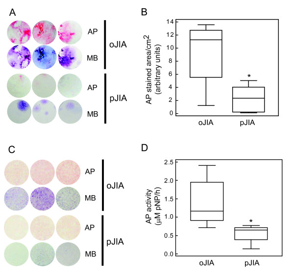
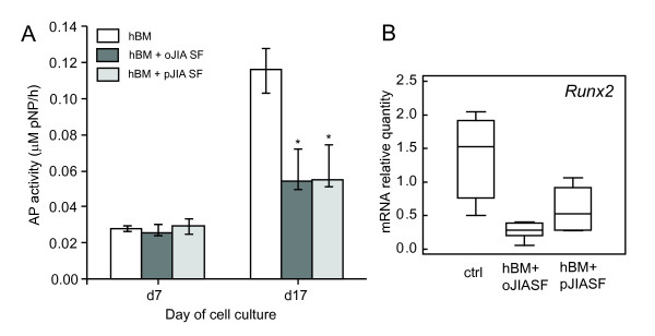
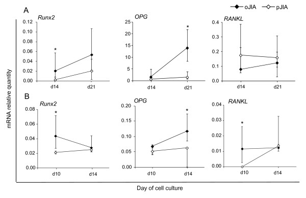
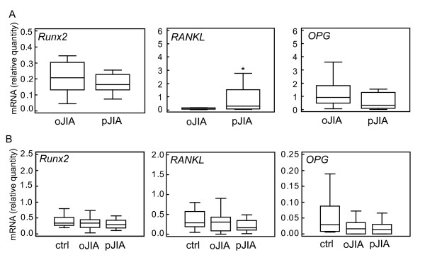
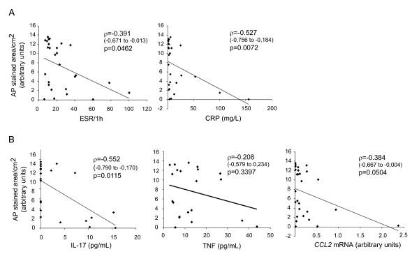
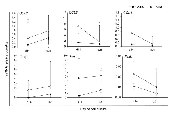
Osteoblastogenesis from SF-derived progenitors was decreased in patients with pJIA compared to those with oJIA. Osteoblastogenesis from primary SF-derived cells negatively correlated with erythrocyte sedimentation rate (ρ = -0.391, P = 0.05), C-reactive protein concentration (ρ = -0.527, P<0.01) and synovial concentration of IL-17 (ρ = -0.552, P = 0.01). SF-derived osteoblasts from pJIA patients expressed more CCL2 and CCL3 genes than in oJIA (P = 0.04 and P = 0.03, respectively; Mann-Whitney test). Expression of Fas was significantly higher in osteoblasts from patients with pJIA than those with oJIA (P = 0.03, Mann-Whitney test). SF-derived cells from patients with pJIA expressed higher levels of RANKL than in oJIA (P = 0.05, Mann-Whitney test). PBMCs from patients with JIA expressed less OPG than healthy control patients (P = 0.05, Kruskal-Wallis test). SF from all tested JIA patients inhibited differentiation of hBM-derived osteoblasts (P = 0.04, Kruskal-Wallis test).
Osteoblast differentiation was decreased in patients with severe forms of JIA and accompanied by altered cytokine/chemokine expression pattern. Development of therapeutic interventions targeting synovial mesenchymal or osteoblast lineage cells in JIA would contribute to alleviating both bone destruction and inflammation in severe forms of the disease.
幼年特发性关节炎(JIA)的特征是滑膜炎症,随后是滑膜的增生性改变,以及关节软骨和下面的骨破坏。这种增生过程是 NF-κB 炎症诱导激活的结果,可能伴随着滑膜间充质祖细胞成骨分化的减少,并有助于骨吸收。我们旨在探讨滑液(SF)来源的间充质祖细胞的成骨分化,并将其与 JIA 患者炎症的严重程度相关联。
收集 18 例寡关节炎(o)JIA 患者、22 例多关节炎(p)JIA 患者和 18 名对照者的外周血,同时收集 18 例 oJIA 患者和 9 例 pJIA 患者的 SF。通过碱性磷酸酶组织化学染色和比色活性测定,评估 SF 来源细胞的成骨分化。评估与成骨相关的基因、Runt 相关转录因子 2(Runx2)、骨保护素(OPG)、核因子 κB 配体受体激活剂(RANKL)和关节炎相关细胞因子/趋化因子基因的表达,肿瘤坏死因子α(TNF-α、Fas、Fas 配体(FasL)、白细胞介素(IL)-1β、IL-4、IL-6、IL-17、IL-18、CC 趋化因子配体(CCL)-2、CCL3、CCL4。将成骨分化与全身和局部炎症指标相关联。还分析了 JIA 患者外周血单核细胞(PBMC)和总 SF 来源细胞中成骨基因的表达。此外,我们评估了 JIA 患者 SF 对人骨髓(hBM)来源成骨细胞分化的抑制作用。
与 oJIA 患者相比,pJIA 患者的 SF 来源祖细胞的成骨分化减少。初级 SF 来源细胞的成骨分化与红细胞沉降率(ρ=-0.391,P=0.05)、C 反应蛋白浓度(ρ=-0.527,P<0.01)和关节 IL-17 浓度(ρ=-0.552,P=0.01)呈负相关。pJIA 患者 SF 来源的成骨细胞表达的 CCL2 和 CCL3 基因多于 oJIA(P=0.04 和 P=0.03,分别为 Mann-Whitney 检验)。与 oJIA 相比,pJIA 患者的成骨细胞中 Fas 的表达显著升高(P=0.03,Mann-Whitney 检验)。pJIA 患者 SF 来源的细胞表达的 RANKL 高于 oJIA(P=0.05,Mann-Whitney 检验)。JIA 患者的 PBMC 表达的 OPG 低于健康对照组患者(P=0.05,Kruskal-Wallis 检验)。来自所有测试 JIA 患者的 SF 抑制了 hBM 来源的成骨细胞的分化(P=0.04,Kruskal-Wallis 检验)。
严重形式的 JIA 患者的成骨分化减少,并伴有细胞因子/趋化因子表达模式的改变。针对 JIA 滑膜间充质或成骨细胞谱系细胞的治疗干预措施的发展,将有助于缓解严重形式疾病的骨破坏和炎症。