Department of Anatomy, Yong Loo Lin School of Medicine, National University of Singapore, Blk MD10, 4 Medical Drive, Singapore, 117597, Singapore.
BMC Neurosci. 2012 Jun 14;13:64. doi: 10.1186/1471-2202-13-64.
Microglia, the resident immune cells of the central nervous system (CNS), have two distinct phenotypes in the developing brain: amoeboid form, known to be amoeboid microglial cells (AMC) and ramified form, known to be ramified microglial cells (RMC). The AMC are characterized by being proliferative, phagocytic and migratory whereas the RMC are quiescent and exhibit a slow turnover rate. The AMC transform into RMC with advancing age, and this transformation is indicative of the gradual shift in the microglial functions. Both AMC and RMC respond to CNS inflammation, and they become hypertrophic when activated by trauma, infection or neurodegenerative stimuli. The molecular mechanisms and functional significance of morphological transformation of microglia during normal development and in disease conditions is not clear. It is hypothesized that AMC and RMC are functionally regulated by a specific set of genes encoding various signaling molecules and transcription factors.
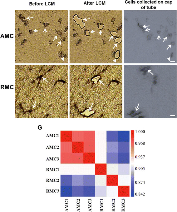
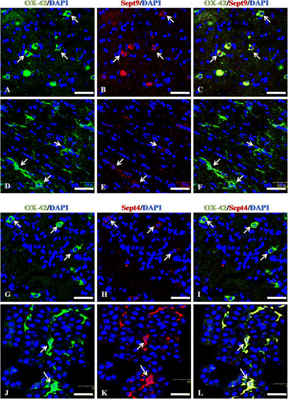
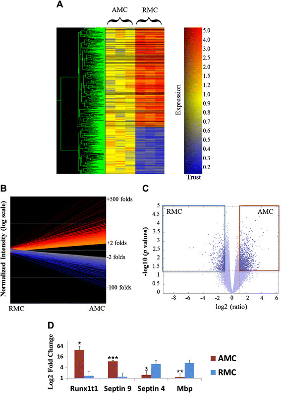
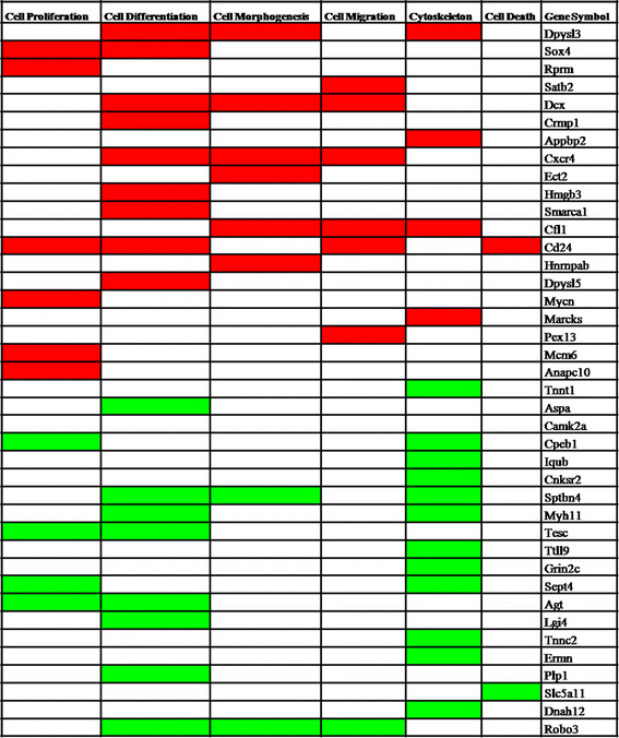
To address this, we carried out cDNA microarray analysis using lectin-labeled AMC and RMC isolated from frozen tissue sections of the corpus callosum of 5-day and 4-week old rat brain respectively, by laser capture microdissection. The global gene expression profiles of both microglial phenotypes were compared and the differentially expressed genes in AMC and RMC were clustered based on their functional annotations. This genome wide comparative analysis identified genes that are specific to AMC and RMC.
The novel and specific molecules identified from the trancriptome explains the quiescent state functioning of microglia in its two distinct morphological states.
小胶质细胞是中枢神经系统(CNS)的固有免疫细胞,在发育中的大脑中有两种截然不同的表型:阿米巴样形态,被称为阿米巴样小胶质细胞(AMC)和分枝状形态,被称为分枝状小胶质细胞(RMC)。AMC 的特征是增殖、吞噬和迁移,而 RMC 则处于静止状态,表现出缓慢的更替率。AMC 随着年龄的增长转化为 RMC,这种转化表明小胶质细胞功能逐渐转变。AMC 和 RMC 均对 CNS 炎症作出反应,当受到创伤、感染或神经退行性刺激激活时,它们会变得肥大。小胶质细胞在正常发育和疾病状态下形态发生转化的分子机制和功能意义尚不清楚。据推测,AMC 和 RMC 受编码各种信号分子和转录因子的特定基因集的功能调节。
为了解决这个问题,我们通过激光捕获显微切割,分别从小鼠大脑胼胝体冷冻组织切片中分离出的 5 天龄和 4 周龄 AMC 和 RMC,使用 lectin 标记,进行了 cDNA 微阵列分析。比较了两种小胶质细胞表型的全基因组基因表达谱,并根据其功能注释对 AMC 和 RMC 中的差异表达基因进行聚类。这项全基因组比较分析确定了 AMC 和 RMC 特有的基因。
从转录组中鉴定出的新的和特异的分子解释了小胶质细胞在两种不同形态状态下的静止功能。