Chaminade Thierry, Marchant Jennifer L, Kilner James, Frith Christopher D
Institut de Neurosciences de la Timone, Campus Santé Timone Marseille, France.
Front Hum Neurosci. 2012 Jun 15;6:179. doi: 10.3389/fnhum.2012.00179. eCollection 2012.
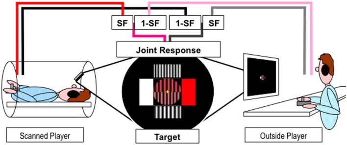
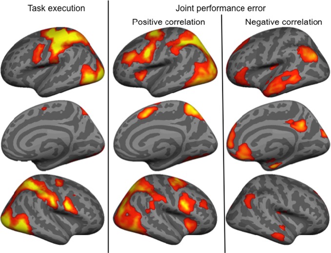
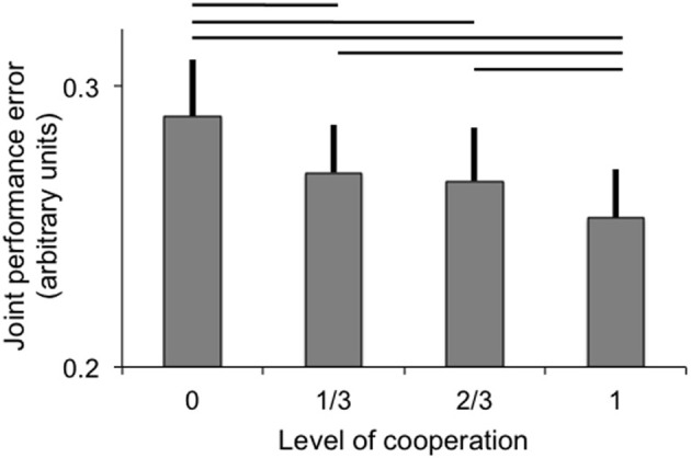
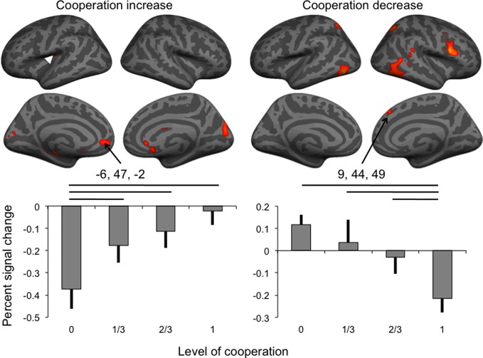
As social agents, humans continually interact with the people around them. Here, motor cooperation was investigated using a paradigm in which pairs of participants, one being scanned with fMRI, jointly controlled a visually presented object with joystick movements. The object oscillated dynamically along two dimensions, color and width of gratings, corresponding to the two cardinal directions of joystick movements. While the overall control of each participant on the object was kept constant, the amount of cooperation along the two dimensions varied along four levels, from no (each participant controlled one dimension exclusively) to full (each participant controlled half of each dimension) cooperation. Increasing cooperation correlated with BOLD signal in the left parietal operculum and anterior cingulate cortex (ACC), while decreasing cooperation correlated with activity in the right inferior frontal and superior temporal gyri, the intraparietal sulci and inferior temporal gyri bilaterally, and the dorsomedial prefrontal cortex. As joint performance improved with the level of cooperation, we assessed the brain responses correlating with behavior, and found that activity in most of the areas associated with levels of cooperation also correlated with the joint performance. The only brain area found exclusively in the negative correlation with cooperation was in the dorso medial frontal cortex, involved in monitoring action outcome. Given the cluster location and condition-related signal change, we propose that this region monitored actions to extract the level of cooperation in order to optimize the joint response. Our results, therefore, indicate that, in the current experimental paradigm involving joint control of a visually presented object with joystick movements, the level of cooperation affected brain networks involved in action control, but not mentalizing.
作为社会主体,人类不断地与周围的人进行互动。在此,我们使用一种范式对运动合作进行了研究,即让参与者两两一组,其中一人接受功能磁共振成像(fMRI)扫描,通过操纵杆移动共同控制一个视觉呈现的物体。该物体沿颜色和光栅宽度这两个维度动态振荡,分别对应操纵杆移动的两个主要方向。虽然每个参与者对物体的总体控制保持不变,但沿这两个维度的合作程度在四个水平上变化,从不合作(每个参与者仅控制一个维度)到完全合作(每个参与者控制每个维度的一半)。合作程度的增加与左侧顶叶岛盖和前扣带回皮质(ACC)的血氧水平依赖(BOLD)信号相关,而合作程度的降低与右侧额下回和颞上回、双侧顶内沟和颞下回以及背内侧前额叶皮质的活动相关。随着联合表现随着合作水平的提高而改善,我们评估了与行为相关的大脑反应,发现与合作水平相关的大多数区域的活动也与联合表现相关。唯一与合作呈负相关的脑区位于背内侧额叶皮质,该区域参与监测行动结果。鉴于簇的位置和与条件相关的信号变化,我们提出该区域监测行动以提取合作水平,从而优化联合反应。因此,我们的结果表明,在当前涉及通过操纵杆移动对视觉呈现物体进行联合控制的实验范式中,合作水平影响参与行动控制的大脑网络,但不影响心理理论。