• 文献检索
  • 文档翻译
  • 深度研究
  • 学术资讯
  • Suppr Zotero 插件Zotero 插件
  • 邀请有礼
  • 套餐&价格
  • 历史记录
应用&插件
Suppr Zotero 插件Zotero 插件浏览器插件Mac 客户端Windows 客户端微信小程序
定价
高级版会员购买积分包购买API积分包
服务
文献检索文档翻译深度研究API 文档MCP 服务
关于我们
关于 Suppr公司介绍联系我们用户协议隐私条款
关注我们

Suppr 超能文献

核心技术专利:CN118964589B侵权必究
粤ICP备2023148730 号-1Suppr @ 2026

文献检索

告别复杂PubMed语法,用中文像聊天一样搜索,搜遍4000万医学文献。AI智能推荐,让科研检索更轻松。

立即免费搜索

文件翻译

保留排版,准确专业,支持PDF/Word/PPT等文件格式,支持 12+语言互译。

免费翻译文档

深度研究

AI帮你快速写综述,25分钟生成高质量综述,智能提取关键信息,辅助科研写作。

立即免费体验

数字化全景X线摄影中下颌骨下缘皮质宽度与形态学之间的相关性评估及绝经后骨质疏松症

Evaluation of Correlation between Width and Morphology of Mandibular Inferior Cortex in Digital Panoramic Radiography and Postmenopausal Osteoporosis.

作者信息

Khojastehpour L, Afsa M, Dabbaghmanesh M H

机构信息

Department of Maxillofacial Radiology, Dental School, Shiraz University of Medical Sciences, Shiraz, Iran.

出版信息

Iran Red Crescent Med J. 2011 Mar;13(3):181-6. Epub 2011 Mar 1.

PMID:22737460
原文链接:https://pmc.ncbi.nlm.nih.gov/articles/PMC3371939/
Abstract

BACKGROUND

In osteoporotic patients, inferior mandibular cortex undergoes resorption which its manifestations can be detected on dental panoramic radiographs as a simple and available method. The aim of this study was to evaluate the correlation between width and morphology of mandibular inferior cortex in digital panoramic radiography and postmenopausal osteoporosis.

METHODS

Bone mineral density (BMD) of lumbar vertebrae and femural neck of 119 postmenopause women was assessed using DXA. Width [cortical index (CI)] and morphology [mandibular cortical index (MCI)] of inferior mandibular cortex were measured and the correlations between BMD and width and shape of the inferior mandibular cortex were evaluated.

RESULTS

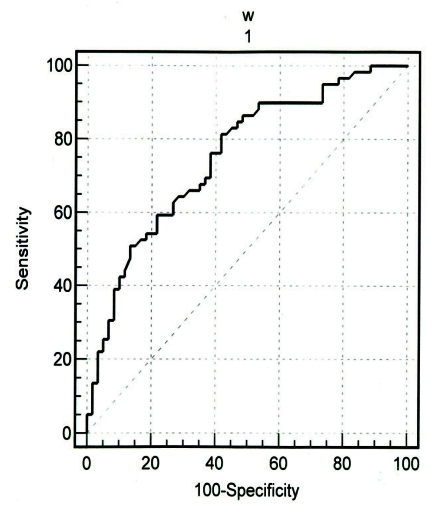
The specificity and sensitivity in identifying women with low BMD of lumbar vertebrae by visual cortical estimation (normal or eroded) were 69.4% and 80.7% respectively. These results in identifying women with low BMD of femural neck were 67.7% and 81.5% respectively. For both sides, the threshold value that provided the highest validity (minimal false negative and false positive results) corresponded to cortical width of 4.29 mm. This threshold in lumbar vertebrae or femural neck provided a sensitivity of 81.4% (95% CI=69.1%-90.3%), specificity of 58.3% (95% CI=44.9%-70.9%), positive predictive value of 65.8% and likelihood ratio of 1.95. There were significant associations between BMD and CI and MCI.

CONCLUSION

Postmenopausal women with thin or eroded mandibular inferior cortex may have an increased risk for low BMD or osteoporosis.

摘要

背景

在骨质疏松患者中,下颌骨下缘皮质会发生吸收,其表现可通过口腔全景X线片检测出来,这是一种简单且可行的方法。本研究的目的是评估数字化全景X线片中下颌骨下缘皮质的宽度和形态与绝经后骨质疏松之间的相关性。

方法

采用双能X线吸收法(DXA)评估119名绝经后女性的腰椎和股骨颈骨密度。测量下颌骨下缘皮质的宽度[皮质指数(CI)]和形态[下颌骨皮质指数(MCI)],并评估骨密度与下颌骨下缘皮质宽度和形态之间的相关性。

结果

通过视觉皮质评估(正常或侵蚀)识别腰椎骨密度低的女性的特异性和敏感性分别为69.4%和80.7%。识别股骨颈骨密度低的女性的这些结果分别为67.7%和81.5%。对于双侧,提供最高有效性(最小假阴性和假阳性结果)的阈值对应于皮质宽度4.29mm。腰椎或股骨颈的这个阈值提供了81.4%(95%CI=69.1%-90.3%)的敏感性、58.3%(95%CI=44.9%-70.9%)的特异性、65.8%的阳性预测值和1.95的似然比。骨密度与CI和MCI之间存在显著关联。

结论

下颌骨下缘皮质薄或有侵蚀的绝经后女性可能有较低骨密度或骨质疏松的风险增加。

https://cdn.ncbi.nlm.nih.gov/pmc/blobs/7122/3371939/c6de234dd2f1/ircmj-13-181-i001.jpg

相似文献

1
Evaluation of Correlation between Width and Morphology of Mandibular Inferior Cortex in Digital Panoramic Radiography and Postmenopausal Osteoporosis.数字化全景X线摄影中下颌骨下缘皮质宽度与形态学之间的相关性评估及绝经后骨质疏松症
Iran Red Crescent Med J. 2011 Mar;13(3):181-6. Epub 2011 Mar 1.
2
Use of dental panoramic radiographs in identifying younger postmenopausal women with osteoporosis.牙科全景X线片在识别年轻绝经后骨质疏松女性中的应用。
Osteoporos Int. 2006;17(3):387-94. doi: 10.1007/s00198-005-2029-7. Epub 2005 Dec 6.
3
Computer-aided system for measuring the mandibular cortical width on dental panoramic radiographs in identifying postmenopausal women with low bone mineral density.用于在牙科全景X线片上测量下颌骨皮质宽度以识别骨质疏松绝经后女性的计算机辅助系统。
Osteoporos Int. 2006;17(5):753-9. doi: 10.1007/s00198-005-0045-2. Epub 2006 Mar 22.
4
Mandibular Inferior Cortex Erosion on Dental Panoramic Radiograph as a Sign of Low Bone Mineral Density in Postmenopausal Women.牙科全景X线片上的下颌骨下缘皮质侵蚀作为绝经后女性低骨密度的标志
Rev Bras Ginecol Obstet. 2017 Dec;39(12):663-669. doi: 10.1055/s-0037-1606622. Epub 2017 Nov 27.
5
Relationship between dental panoramic radiographic findings and biochemical markers of bone turnover.口腔全景X线片表现与骨转换生化标志物之间的关系。
J Bone Miner Res. 2003 Sep;18(9):1689-94. doi: 10.1359/jbmr.2003.18.9.1689.
6
Dental panoramic radiograph as a tool to detect postmenopausal women with low bone mineral density: untrained general dental practitioners' diagnostic performance.牙科全景X线片作为检测绝经后低骨密度女性的工具:未经培训的普通牙科医生的诊断性能
Osteoporos Int. 2003 Aug;14(8):659-64. doi: 10.1007/s00198-003-1419-y. Epub 2003 Jun 24.
7
Digital panoramic radiographs are useful for diagnosis of osteoporosis in Korean postmenopausal women.数字化全景X线片对韩国绝经后女性骨质疏松症的诊断很有用。
Gerodontology. 2016 Jun;33(2):185-92. doi: 10.1111/ger.12134. Epub 2014 Jun 9.
8
Panoramic-based mandibular indices in relation to mandibular bone mineral density and skeletal status assessed by dual energy X-ray absorptiometry and quantitative ultrasound.基于全景片的下颌骨指数与通过双能X线吸收法和定量超声评估的下颌骨骨密度及骨骼状态的关系
Dentomaxillofac Radiol. 2002 Nov;31(6):361-7. doi: 10.1038/sj.dmfr.4600729.
9
[Estimation of mandibular bone status and lumbar bone mineral density in postmenopausal women].绝经后女性下颌骨状况及腰椎骨密度的评估
Pol Merkur Lekarski. 2016 Aug;41(242):79-83.
10
Visual assessment of the mandibular cortex on panoramic radiographs to identify postmenopausal women with low bone mineral densities.通过全景X线片对下颌骨皮质进行视觉评估,以识别骨矿物质密度低的绝经后女性。
Oral Surg Oral Med Oral Pathol Oral Radiol Endod. 2005 Aug;100(2):226-31. doi: 10.1016/j.tripleo.2004.11.052.

引用本文的文献

1
Assessment of the efficacy of digital panoramic radiographs in analyzing changes in bone mineral density in postmenopausal women.评估数字化全景X线片在分析绝经后女性骨密度变化中的效能。
J Family Med Prim Care. 2022 Aug;11(8):4342-4348. doi: 10.4103/jfmpc.jfmpc_13_22. Epub 2022 Aug 30.
2
Clinical guidelines for the application of panoramic radiographs in screening for osteoporosis.临床应用全景 X 光片筛查骨质疏松症指南。
Oral Radiol. 2021 Apr;37(2):189-208. doi: 10.1007/s11282-021-00518-6. Epub 2021 Feb 23.
3
Mandibular cortical index in the screening of postmenopausal at low mineral density risk: a systematic review.

本文引用的文献

1
Dental and radiographic findings as predictors of osteoporosis in postmenopausal women.牙科及影像学检查结果作为绝经后女性骨质疏松症的预测指标
Geriatr Gerontol Int. 2009 Jun;9(2):155-64. doi: 10.1111/j.1447-0594.2009.00518.x.
2
Detection of post-menopausal women with low bone mineral density and elevated biochemical markers of bone turnover by panoramic radiographs.通过全景X线片检测绝经后骨密度低且骨转换生化标志物升高的女性。
Dentomaxillofac Radiol. 2008 Dec;37(8):433-7. doi: 10.1259/dmfr/85235532.
3
Assessment of panoramic radiomorphometric indices in Turkish patients of different age groups, gender and dental status.
下颌骨皮质指数在低骨密度风险绝经后妇女筛查中的应用:系统评价。
Dentomaxillofac Radiol. 2021 May 1;50(4):20200514. doi: 10.1259/dmfr.20200514. Epub 2021 Feb 17.
4
Relationship between Two Panoramic Radiography Indices and Bone Mineral Density of Postmenopausal Women with Osteopenia and Osteoporosis.两种全景X线摄影指标与骨质疏松症和骨质减少绝经后女性骨密度的关系
J Dent (Shiraz). 2018 Sep;19(3):181-188.
5
Panoramic radiographs and quantitative ultrasound of the radius and phalanx III to assess bone mineral status in postmenopausal women.使用全景X线片以及第三掌骨和桡骨的定量超声评估绝经后女性的骨矿物质状况。
BMC Oral Health. 2018 Jul 28;18(1):127. doi: 10.1186/s12903-018-0593-4.
6
Panoramic radiographs for detecting osteopenia: A pilot study.用于检测骨质减少的全景X线片:一项初步研究。
Clin Pract. 2017 Oct 19;7(4):973. doi: 10.4081/cp.2017.973. eCollection 2017 Sep 15.
7
IDIOS: An innovative index for evaluating dental imaging-based osteoporosis screening indices.IDIOS:一种用于评估基于牙科影像的骨质疏松症筛查指标的创新指数。
Imaging Sci Dent. 2016 Sep;46(3):185-202. doi: 10.5624/isd.2016.46.3.185. Epub 2016 Sep 20.
8
Feasibility of cone beam computed tomography radiomorphometric analysis and fractal dimension in assessment of postmenopausal osteoporosis in correlation with dual X-ray absorptiometry.锥形束计算机断层扫描放射形态计量学分析及分形维数在评估绝经后骨质疏松症与双能X线吸收法相关性中的可行性。
Dentomaxillofac Radiol. 2016;45(7):20160212. doi: 10.1259/dmfr.20160212. Epub 2016 Aug 2.
9
Can dental pulp calcification predict the risk of ischemic cardiovascular disease?牙髓钙化能否预测缺血性心血管疾病的风险?
J Dent (Tehran). 2013 Sep;10(5):456-60. Epub 2013 Sep 30.
10
Prediction of lumbar spine bone mineral density from the mandibular cortical width in postmenopausal women.根据绝经后女性下颌骨皮质宽度预测腰椎骨密度
J Res Med Sci. 2013 Nov;18(11):951-5.
不同年龄组、性别和牙齿状况的土耳其患者全景放射形态计量学指标评估。
Dentomaxillofac Radiol. 2008 Jul;37(5):288-92. doi: 10.1259/dmfr/19491030.
4
Observer performance in diagnosing osteoporosis by dental panoramic radiographs: results from the osteoporosis screening project in dentistry (OSPD).通过牙科全景X线片诊断骨质疏松症时观察者的表现:牙科骨质疏松症筛查项目(OSPD)的结果
Bone. 2008 Jul;43(1):209-213. doi: 10.1016/j.bone.2008.03.014. Epub 2008 Apr 8.
5
Mandibular radiomorphometric measurements as indicators of possible osteoporosis in postmenopausal women.下颌骨放射形态测量作为绝经后女性骨质疏松症可能性的指标。
Maturitas. 2007 Nov 20;58(3):226-35. doi: 10.1016/j.maturitas.2007.08.014. Epub 2007 Oct 17.
6
Risk of vertebral osteoporosis in post-menopausal women with alterations of the mandible.下颌骨改变的绝经后女性患椎体骨质疏松症的风险。
Dentomaxillofac Radiol. 2007 Mar;36(3):143-8. doi: 10.1259/dmfr/50171930.
7
The differences in panoramic mandibular indices and fractal dimension between patients with and without spinal osteoporosis.患有和未患有脊柱骨质疏松症的患者之间全景下颌指数和分形维数的差异。
Dentomaxillofac Radiol. 2006 Jan;35(1):1-9. doi: 10.1259/dmfr/97652136.
8
Use of dental panoramic radiographs in identifying younger postmenopausal women with osteoporosis.牙科全景X线片在识别年轻绝经后骨质疏松女性中的应用。
Osteoporos Int. 2006;17(3):387-94. doi: 10.1007/s00198-005-2029-7. Epub 2005 Dec 6.
9
Visual assessment of the mandibular cortex on panoramic radiographs to identify postmenopausal women with low bone mineral densities.通过全景X线片对下颌骨皮质进行视觉评估,以识别骨矿物质密度低的绝经后女性。
Oral Surg Oral Med Oral Pathol Oral Radiol Endod. 2005 Aug;100(2):226-31. doi: 10.1016/j.tripleo.2004.11.052.
10
Radiomorphometric indices and their relation to gender, age, and dental status.放射形态计量学指标及其与性别、年龄和牙齿状况的关系。
Oral Surg Oral Med Oral Pathol Oral Radiol Endod. 2005 Apr;99(4):479-84. doi: 10.1016/j.tripleo.2004.09.013.