Institute of Cell Biology, Zhejiang University, Hangzhou 310058, China.
Int J Biol Sci. 2012;8(6):901-12. doi: 10.7150/ijbs.4554. Epub 2012 Jun 22.
Oridonin (ORI) could inhibit the proliferation and induce apoptosis in various cancer cell lines. However, the mechanism is not fully understood.
Human prostate cancer (HPC) cells were cultured in vitro and cell viability was detected by the CCK-8 assay. The ultrastructure changes were observed under transmission electron microscope (TEM). Chemical staining with acridine orange (AO), MDC or DAPI was used to detect acidic vesicular organelles (AVOs) and alternation of DNA. Expression of LC3 and P21 was detected by Western Blot. Apoptotic rates and cell cycle arrest were detected by FACS.
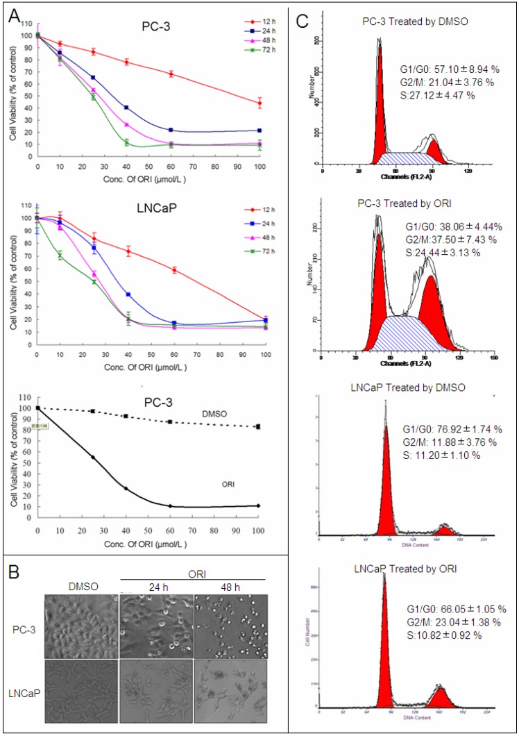
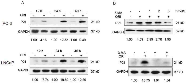
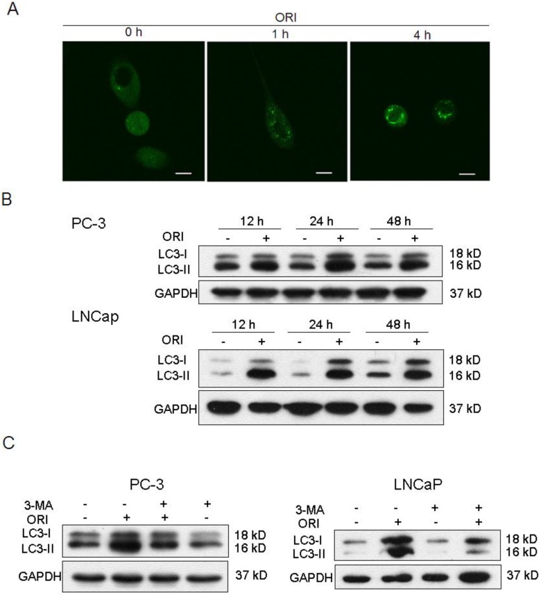
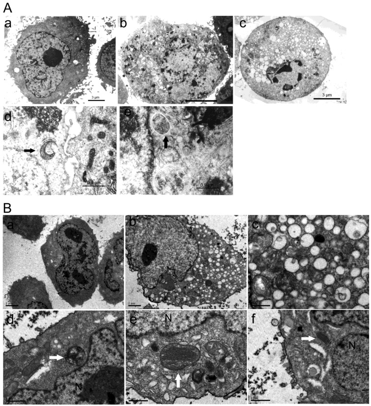
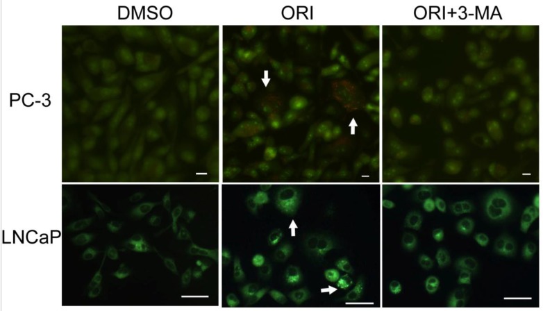
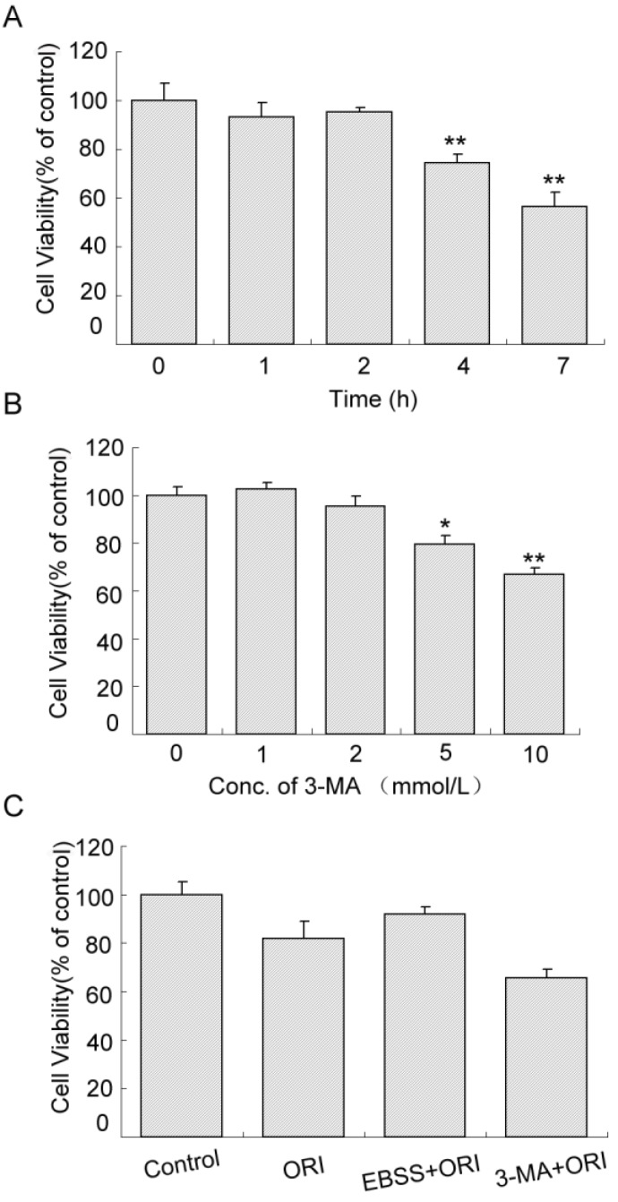
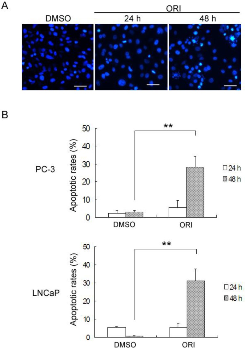
Our study demonstrated that after ORI treatment, the proliferations of human prostate cancer (HPC) cell lines PC-3 and LNCaP were inhibited in a concentration and time-dependent manner. ORI induced cell cycle arrest at the G2/M phase. A large number of autophagosomes with double-membrane structure and acidic vesicular organelles (AVOs) were detected in the cytoplasm of HPC cells treated with ORI for 24 hours. ORI resulted in the conversion of LC3-I to LC3-II and recruitment of LC3-II to the autophagosomal membranes. Autophagy inhibitor 3-methyladenine (3-MA) reduced AVOs formation and inhibited LC3-I to LC3-II conversion. At 48 h, DNA fragmentation, chromatin condensation and disappearance of surface microvilli were detected in ORI-treated cells. ORI induced a significant increase in the number of apoptotic cells (PC-3: 5.4% to 27.0%, LNCaP: 5.3% to 31.0%). Promoting autophagy by nutrient starvation increased cell viability, while inhibition of autophagy by 3-MA promoted cell death. The expression of P21 was increased by ORI, which could be completely reversed by the inhibition of autophagy.
Our findings indicated that autophagy occurred before the onset of apoptosis and protected cancer cells in ORI-treated HPC cells. P21 was involved in ORI-induced autophagy and apoptosis. Our results provide an experimental basis for understand the anti-tumor mechanism of ORI as treatment for prostate cancer.
冬凌草甲素(ORI)可抑制多种癌细胞系的增殖并诱导细胞凋亡。然而,其机制尚不完全清楚。
体外培养人前列腺癌细胞(HPC),用 CCK-8 法检测细胞活力。用透射电子显微镜(TEM)观察超微结构变化。用吖啶橙(AO)、MDC 或 DAPI 化学染色检测酸性囊泡细胞器(AVOs)和 DNA 改变。用 Western Blot 检测 LC3 和 P21 的表达。用 FACS 检测细胞凋亡率和细胞周期阻滞。
本研究表明,ORI 处理后,人前列腺癌细胞系 PC-3 和 LNCaP 的增殖呈浓度和时间依赖性抑制。ORI 诱导细胞周期阻滞于 G2/M 期。在 ORI 处理 24 小时的 HPC 细胞的细胞质中,检测到大量具有双层膜结构和酸性囊泡细胞器(AVOs)的自噬体。ORI 导致 LC3-I 转化为 LC3-II,并募集 LC3-II 到自噬体膜上。自噬抑制剂 3-甲基腺嘌呤(3-MA)减少 AVOs 形成并抑制 LC3-I 转化为 LC3-II。在 48 小时时,在 ORI 处理的细胞中检测到 DNA 片段化、染色质浓缩和表面微绒毛消失。ORI 诱导凋亡细胞数量显著增加(PC-3:5.4%至 27.0%,LNCaP:5.3%至 31.0%)。营养饥饿促进自噬可增加细胞活力,而 3-MA 抑制自噬可促进细胞死亡。ORI 增加 P21 的表达,该表达可被自噬抑制完全逆转。
我们的研究结果表明,自噬发生在凋亡开始之前,并在 ORI 处理的 HPC 细胞中保护癌细胞。P21 参与 ORI 诱导的自噬和凋亡。我们的结果为理解 ORI 作为前列腺癌治疗药物的抗肿瘤机制提供了实验依据。