Department of Pharmacology and Physiology, Saint Louis University, 1402 South Grand Blvd, St. Louis, MO 63104, USA.
J Neuroinflammation. 2012 Jun 29;9:150. doi: 10.1186/1742-2094-9-150.
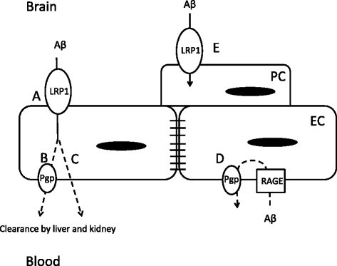
Defects in the low density lipoprotein receptor-related protein-1 (LRP-1) and p-glycoprotein (Pgp) clearance of amyloid beta (Aβ) from brain are thought to contribute to Alzheimer's disease (AD). We have recently shown that induction of systemic inflammation by lipopolysaccharide (LPS) results in impaired efflux of Aβ from the brain. The same treatment also impairs Pgp function. Here, our aim is to determine which physiological routes of Aβ clearance are affected following systemic inflammation, including those relying on LRP-1 and Pgp function at the blood-brain barrier.
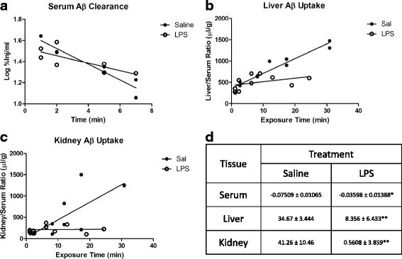
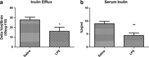
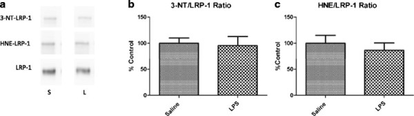
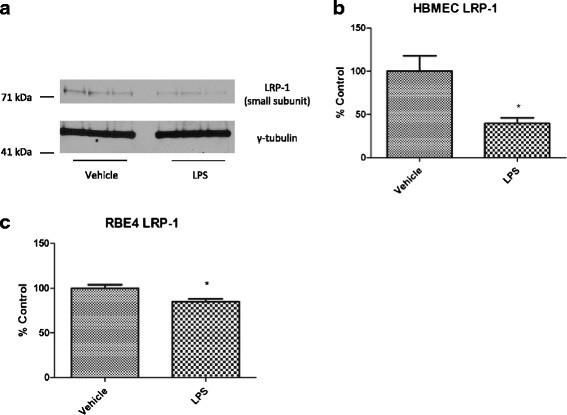
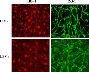
CD-1 mice aged between 6 and 8 weeks were treated with 3 intraperitoneal injections of 3 mg/kg LPS at 0, 6, and 24 hours and studied at 28 hours. 125I-Aβ1-42 or 125I-alpha-2-macroglobulin injected into the lateral ventricle of the brain (intracerebroventricular (ICV)) or into the jugular vein (intravenous (IV)) was used to quantify LRP-1-dependent partitioning between the brain vasculature and parenchyma and peripheral clearance, respectively. Disappearance of ICV-injected 14 C-inulin from brain was measured to quantify bulk flow of cerebrospinal fluid (CSF). Brain microvascular protein expression of LRP-1 and Pgp was measured by immunoblotting. Endothelial cell localization of LRP-1 was measured by immunofluorescence microscopy. Oxidative modifications to LRP-1 at the brain microvasculature were measured by immunoprecipitation of LRP-1 followed by immunoblotting for 4-hydroxynonenal and 3-nitrotyrosine.
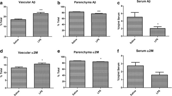
We found that LPS: caused an LRP-1-dependent redistribution of ICV-injected Aβ from brain parenchyma to brain vasculature and decreased entry into blood; impaired peripheral clearance of IV-injected Aβ; inhibited reabsorption of CSF; did not significantly alter brain microvascular protein levels of LRP-1 or Pgp, or oxidative modifications to LRP-1; and downregulated LRP-1 protein levels and caused LRP-1 mislocalization in cultured brain endothelial cells.
These results suggest that LRP-1 undergoes complex functional regulation following systemic inflammation which may depend on cell type, subcellular location, and post-translational modifications. Our findings that systemic inflammation causes deficits in both Aβ transport and bulk flow like those observed in AD indicate that inflammation could induce and promote the disease.
载脂蛋白β(Aβ)从大脑中的 LDL 受体相关蛋白-1(LRP-1)和 p-糖蛋白(Pgp)清除缺陷被认为是导致阿尔茨海默病(AD)的原因。我们最近发现,脂多糖(LPS)诱导的全身炎症会导致 Aβ从大脑中的流出受损。同样的治疗也会损害 Pgp 的功能。在这里,我们的目的是确定全身炎症后哪些 Aβ清除的生理途径受到影响,包括依赖于血脑屏障上的 LRP-1 和 Pgp 功能的途径。
6 至 8 周龄的 CD-1 小鼠接受 3 次腹腔内注射 3mg/kg LPS,分别在 0、6 和 24 小时进行,然后在 28 小时进行研究。125I-Aβ1-42 或 125I-α-2-巨球蛋白注入侧脑室(脑室内(ICV))或颈静脉(静脉内(IV))用于分别定量 LRP-1 依赖性分配到大脑血管和实质之间以及外周清除率。通过测量脑室内注射的 14C- 尿囊素从大脑中的消失来定量脑脊液(CSF)的总体流动。通过免疫印迹测量 LRP-1 和 Pgp 的脑微血管蛋白表达。通过免疫荧光显微镜测量 LRP-1 在脑微血管内皮细胞中的定位。通过免疫沉淀 LRP-1 后进行免疫印迹测量 4-羟基壬烯醛和 3-硝基酪氨酸来测量 LRP-1 在脑微血管上的氧化修饰。
我们发现 LPS:导致 ICV 注射的 Aβ从大脑实质向大脑血管的 LRP-1 依赖性重新分布,并减少进入血液;损害 IV 注射的 Aβ的外周清除率;抑制 CSF 的重吸收;对脑微血管 LRP-1 或 Pgp 的蛋白水平或 LRP-1 的氧化修饰没有显著影响;并下调 LRP-1 蛋白水平并导致培养的脑内皮细胞中 LRP-1 定位错误。
这些结果表明,LRP-1 在全身炎症后经历复杂的功能调节,这可能取决于细胞类型、亚细胞位置和翻译后修饰。我们发现全身炎症会导致 Aβ转运和总体流动的缺陷,类似于 AD 中观察到的缺陷,表明炎症可能会引发和促进疾病。