Koestenberger Martin
Division of Pediatric Cardiology, Department of Pediatrics, Medical University of Graz, Auenbruggerplatz 30, 8036 Graz, Austria.
ISRN Pediatr. 2012;2012:753481. doi: 10.5402/2012/753481. Epub 2012 Jun 13.
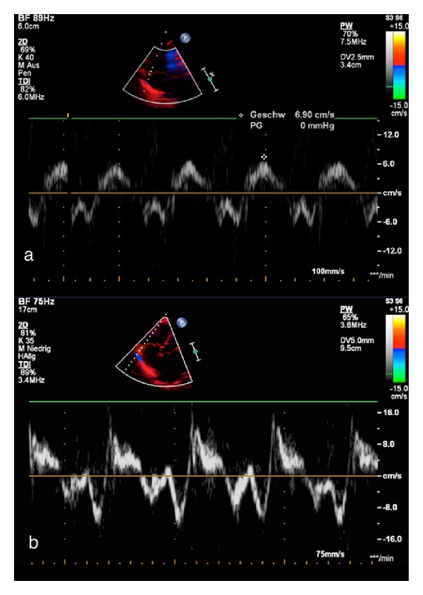
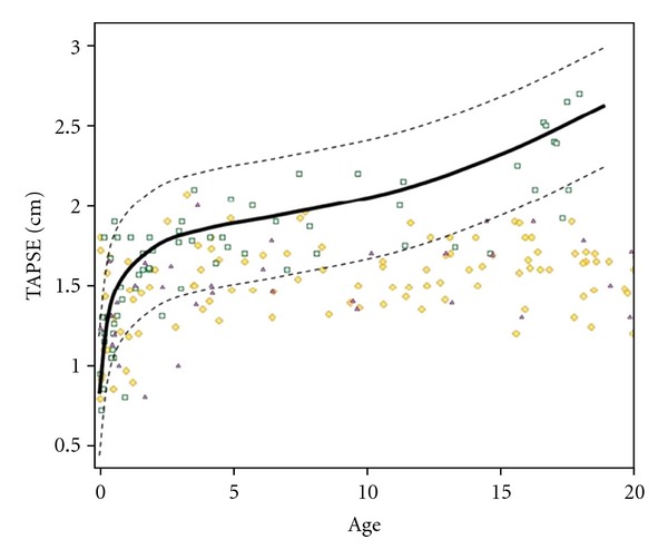
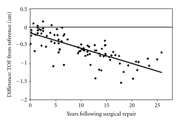
Transthoracic echocardiography (TTE) is the first-line tool for diagnosis and followup of pediatric and young adult patients with congenital heart disease (CHD). Appropriate use of TTE can reduce the need for more invasive modalities, such as cardiac catheterization and cardiac magnetic resonance imaging. New echocardiographic techniques have emerged more recently: tissue Doppler imaging, tissue tracking (strain and strain rate), vector velocity imaging (VVI), myocardial performance index, myocardial acceleration during isovolumic acceleration (IVA), the ratio of systolic to diastolic duration (S/D ratio), and two dimensional measurements of systolic right ventricular (RV) function (e.g., tricuspid annular plane systolic excursion, TAPSE). These may become valuable indicators of ventricular performance, compliance, and disease progression. In addition, three-dimensional (3D) echocardiography when performed for the assessment of valvular function, device position, and ventricular volumes is being integrated into routine clinical care. In this paper, the potential use and limitations of these new echocardiographic techniques in patients with CHD are discussed. A particular focus is on the echocardiographic assessment of right ventricular (RV) function in conditions associated with increased right ventricular volume (e.g., pulmonary regurgitation after tetralogy of Fallot repair) or pressure (e.g., pulmonary hypertension) in children and young adults.
经胸超声心动图(TTE)是先天性心脏病(CHD)儿科及青年患者诊断和随访的一线工具。合理使用TTE可减少对侵入性更强的检查手段的需求,如心导管检查和心脏磁共振成像。最近出现了一些新的超声心动图技术:组织多普勒成像、组织追踪(应变和应变率)、向量速度成像(VVI)、心肌性能指数、等容加速期心肌加速度(IVA)、收缩期与舒张期时长比值(S/D比值)以及右心室(RV)收缩功能的二维测量(如三尖瓣环平面收缩期位移,TAPSE)。这些可能成为心室功能、顺应性和疾病进展的重要指标。此外,用于评估瓣膜功能、装置位置和心室容积的三维(3D)超声心动图正被纳入常规临床护理。本文讨论了这些新超声心动图技术在CHD患者中的潜在用途和局限性。特别关注儿童和青年患者在右心室容积增加(如法洛四联症修复术后肺动脉反流)或压力升高(如肺动脉高压)情况下右心室(RV)功能的超声心动图评估。