Czarnecka Anna, Zimny Anna, Sąsiadek Marek
Department of General Radiology, Interventional Radiology and Neuroradiology, Chair of Radiology, Wrocław Medical University, Wrocław, Poland.
Pol J Radiol. 2010 Apr;75(2):15-21.
Both brain atrophy and decrease of perfusion are observed in dementive diseases. The aim of the study was to correlate the results of brain perfusion CT (pCT) and CT volumetry in patients with Alzheimer's disease (AD).
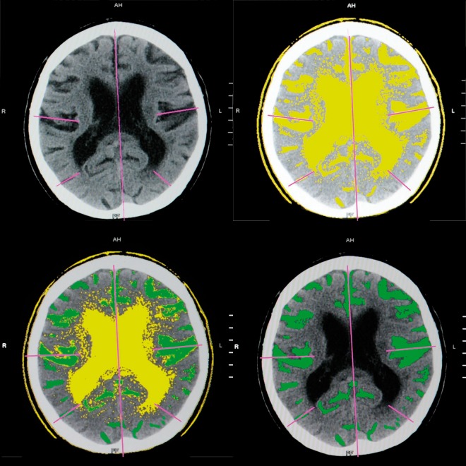
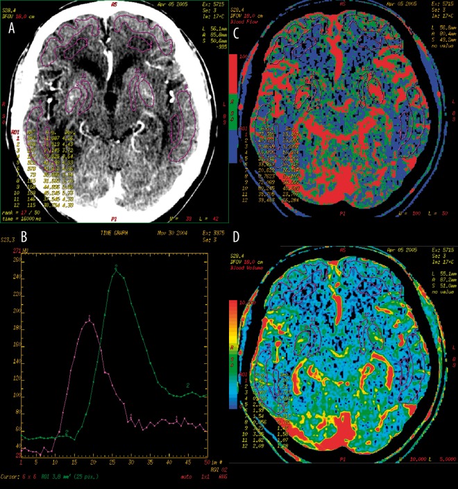
MATERIAL/METHODS: Forty-eight patients with AD (mean age of 71.3 years) underwent brain pCT and CT volumetry. The pCT was performed at the level of basal ganglia after the injection of contrast medium (50 ml, 4 ml/sec.) with serial scanning (delay 7 sec, 50 scans, 1 scan/sec). Volumetric measurements were carried out on the basis of source images, with the use of a dedicated CT software combined with manual outlining of the regions of interest in extracerebral and intraventricular CSF spaces. Perfusion parameters of the cerebral blood flow (CBF) and cerebral blood volume (CBV) from the grey matter of frontal and temporal as well as basal ganglia were compared statistically with the volumetric measurements of frontal and temporal cortical atrophy as well as subcortical atrophy.
A statistically significant positive correlation was found between the values of CBF and CBV in the basal ganglia and the volumes of the lateral and third ventricles. The comparison of CBF and CBV results with the volumetric measurements in the areas of the frontal and temporal lobes showed mostly negative correlations, but none of them was of statistical significance.
In patients with AD, the degree of cortical atrophy is not correlated with the decrease of perfusion in the grey matter and subcortical atrophy is not correlated with the decrease of perfusion in the basal ganglia region. It suggests that functional and structural changes in AD are not related to each other.
在痴呆性疾病中可观察到脑萎缩和灌注降低。本研究的目的是将阿尔茨海默病(AD)患者的脑灌注CT(pCT)结果与CT容积测量结果进行相关性分析。
材料/方法:48例AD患者(平均年龄71.3岁)接受了脑pCT和CT容积测量。在注射造影剂(50ml,4ml/秒)后,于基底节水平进行pCT检查,并进行连续扫描(延迟7秒,50次扫描,每秒1次扫描)。基于源图像进行容积测量,使用专用的CT软件并结合手动勾勒脑外和脑室内脑脊液间隙的感兴趣区域。将额叶、颞叶以及基底节灰质的脑血流量(CBF)和脑血容量(CBV)灌注参数与额叶和颞叶皮质萎缩以及皮质下萎缩的容积测量结果进行统计学比较。
基底节区的CBF和CBV值与侧脑室和第三脑室的容积之间存在统计学上显著的正相关。CBF和CBV结果与额叶和颞叶区域的容积测量结果比较大多呈负相关,但均无统计学意义。
在AD患者中,皮质萎缩程度与灰质灌注降低无关,皮质下萎缩与基底节区灌注降低无关。这表明AD中的功能和结构变化彼此无关。