Michalak Maciej, Zurada Anna, Biernacki Maciej, Zygmunt Kozielec
Department of Oncology, Faculty of Medical Sciences, University of Warmia and Mazury in Olsztyn, Olsztyn, Poland.
Pol J Radiol. 2010 Oct;75(4):44-6.
The rupture of ectopic pregnancy (EP) still remains the primary and direct cause of death in the first trimester of pregnancy. Ultrasonography is known to be a modality of choice in EP diagnostics. We found a severe discrepancy between the frequency of ectopic pregnancies (EP) and the number of available computed tomography (CT) examinations.
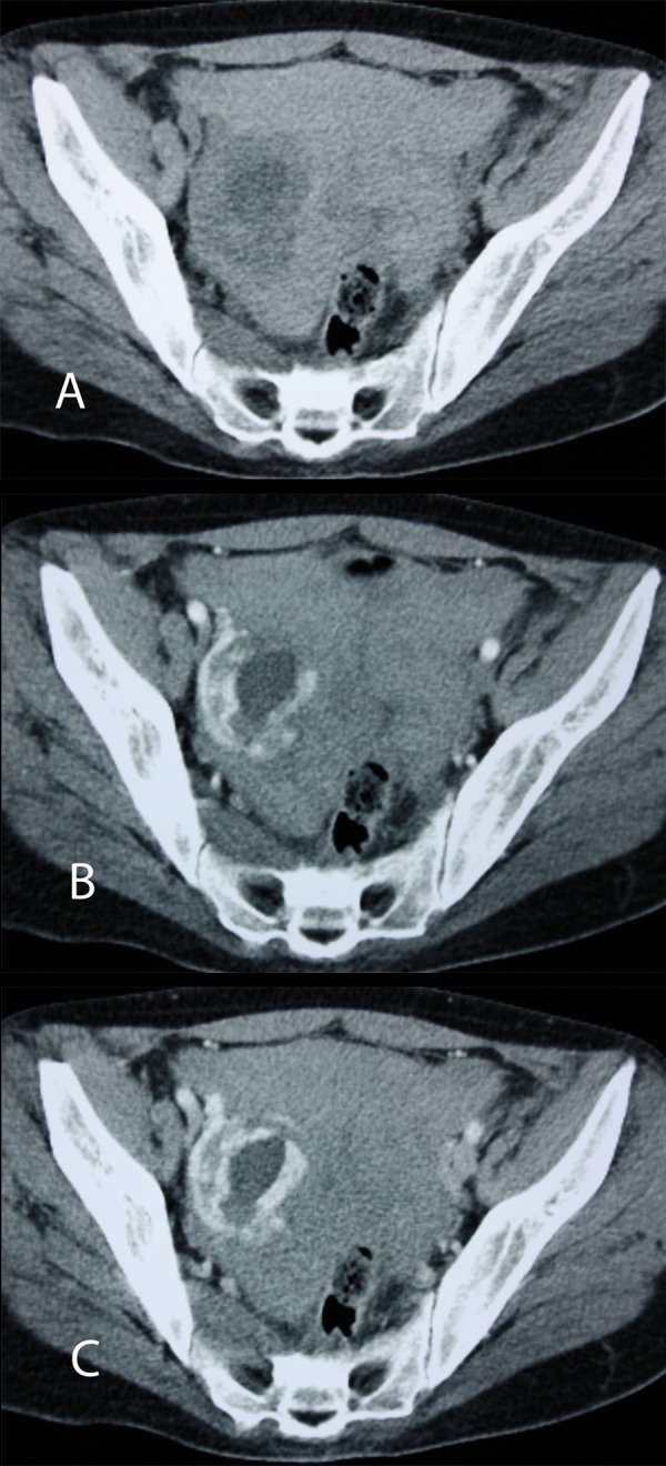
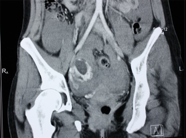
A 29-year-old woman was admitted to the emergency department with a history of abdominal pain, nausea, vomiting and collapse. Sonographic findings of a suspected EP were unclear. Moreover, not all features of intrauterine pregnancy were present. Due to the patient's life-threatening condition, an emergency multi-slice CT with MPR and VRT reconstructions was performed, revealing symptoms of a ruptured EP. In the right adnexal area, a well-vascularized, solid-cystic abnormal mass lesion was found. Intraperitoneal hemorrhage was confirmed intraoperatively, and the right fallopian tube with a tubal EP was resected. In the surgery in situ, as well as in the pathological examination of the tumor mass, a human embryo of approximately 1.5 cm in length (beginning of the 8(th) week of gestation) was found.
Although ultrasonography still remains the first-line imaging examination in EP diagnostics, sometimes the findings of suspected EPs are unclear and not sufficient. The rupture of EP, with serious bleeding and symptoms of shock, may require an emergent pelvic and abdominal CT inspection. A clear correlation was found between the macroscopic CT images and the intraoperatively sampled material.
异位妊娠(EP)破裂仍然是妊娠早期死亡的主要直接原因。超声检查是诊断EP的首选方法。我们发现异位妊娠(EP)的发生率与计算机断层扫描(CT)检查的可用数量之间存在严重差异。
一名29岁女性因腹痛、恶心、呕吐和晕厥病史被收入急诊科。疑似EP的超声检查结果不明确。此外,宫内妊娠的所有特征均未出现。由于患者生命垂危,进行了一次带有多平面重组(MPR)和容积再现技术(VRT)重建的急诊多层CT检查,结果显示为EP破裂的症状。在右侧附件区发现一个血运丰富的实性囊性异常肿块病变。术中证实有腹腔内出血,并切除了右侧输卵管及输卵管妊娠物。在原位手术以及肿瘤肿块的病理检查中,发现了一个长约1.5厘米(妊娠第8周开始)的人类胚胎。
尽管超声检查仍然是诊断EP的一线影像学检查方法,但有时疑似EP的检查结果不明确且不充分。EP破裂伴有严重出血和休克症状时,可能需要紧急进行盆腔和腹部CT检查。在宏观CT图像与术中取样材料之间发现了明确的相关性。